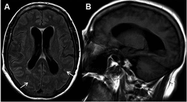
Figure 3. JC Virus Meningitis (JCVM).
MRI demonstrates hydrocephalus, abnormal signal in subarachnoid space, and no parenchymal brain lesions. (A) Axial fluid-attenuated inversion recovery sequence shows enlarged ventricles and abnormal hyperintensity in the subarachnoid space, within the sulci of the cerebral hemispheres (arrows). (B) A sagittal T1-weighted sequence demonstrates significant enlargement of the lateral ventricle.

