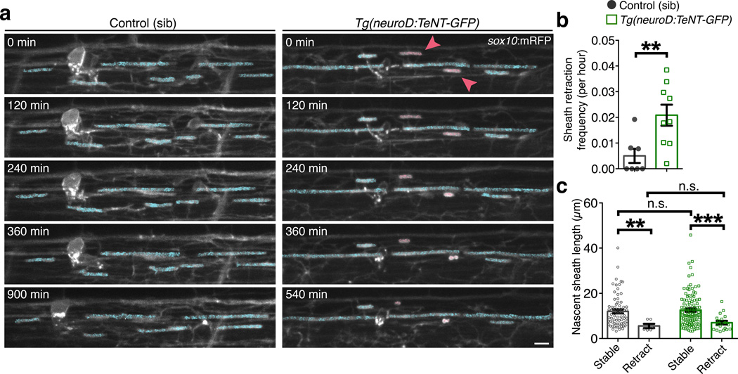
Figure 6. Nascent myelin sheaths are stabilized by activity-dependent secretion.
(a) Representative confocal images show the retraction of existing sheaths during 15-hr time-lapse imaging in sibling control and Tg(neuroD:TeNT-GFP) larvae. Images are lateral views of the dorsal spinal cord and the time (min) relative to the start of image acquisition is indicated for each image. For demonstrative purposes, sheaths stable for the entire time-lapse are shaded in blue and retracting sheaths are shaded red. Scale bar, 5 µm. (b) Summary of time-lapse measurements show the frequency of sheath retraction. n = 7 control (99 sheaths) and 9 Tg(neuroD:TeNT-GFP) larvae (129 sheaths). P = 0.0071, Mann-Whitney test. (c) Quantitative measurements show the relationship between sheath length and stability. Bars represent the mean ± s.e.m. n = 7 control larvae (72 stable sheaths, 6 retracting sheaths) and 9 Tg(neuroD:TeNT-GFP) larvae (106 stable sheaths, 19 retracting sheaths). P = 0.0034 (left, **), P = 0.0002 (right, ***), P = 0.3737 (upper n.s.), P = 0.6105 (lower n.s.), Mann-Whitney test; For (b–c), **P < 0.01, **P < 0.001.

