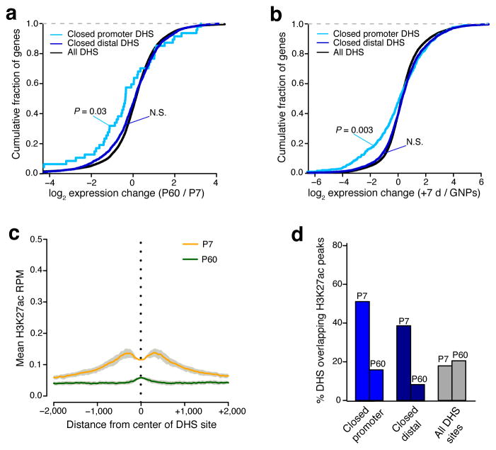
Figure 4. A subset of closing DHS sites mark early postnatal enhancer elements.
(a,b) Cumulative fraction of genes nearest to closed promoter-located, closed distal, and all identified DHS sites with given fold-change in RNA-seq expression from P7 to P60 cerebellum (a) or from GNPs to +7DIV (b). Leftward shift indicates genes decreased in expression across developmental time. Significance assessed by Mann-Whitney U test. (c) Mean H3K27ac ChIP-seq signal (reads per million mapped) present at center of P7 to P60 closed DHS sites (5503 sites) in either P7 cerebellum (orange line) or P60 cerebellum (green line). Gray = s.e.m. (n = 2 biological replicates of pooled cerebella). (d) Percent of closed promoter-located, closed distal, and all DHS sites overlapping H3K27ac peaks identified in P7 or P60 cerebellum.

