
Fig. 6.
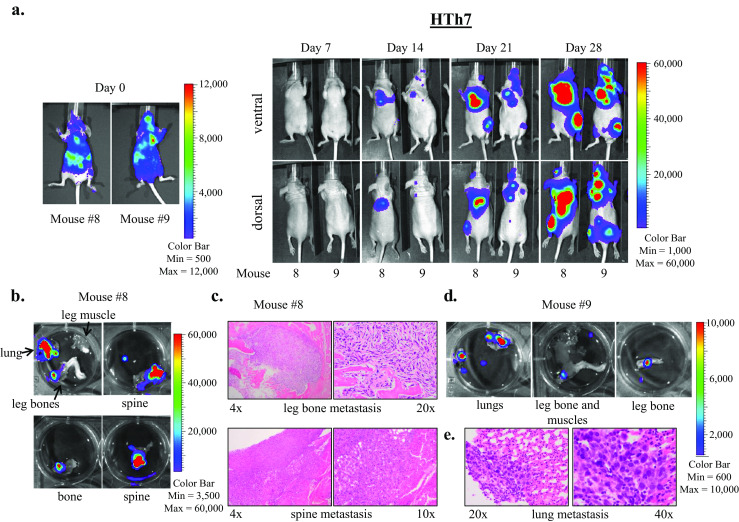
HTh7 cells form widespread metastases to lungs, bone, and brain in the intracardiac metastasis model. Images of two representative mice imaged weekly using the Xenogen IVIS 200 imaging system after intracardiac injection of HTh7 cells are shown (a). Ex vivo IVIS imaging reveals metastases to lungs and bone for mouse 8 (b) and mouse 9 (d), and metastases were confirmed by histologic examination. Bone metastases to the leg bone and spine are shown at 4×, 10×, and 20× magnification (c), and lung metastasis from mouse 9 is shown at ×20 and ×40 magnification (e)