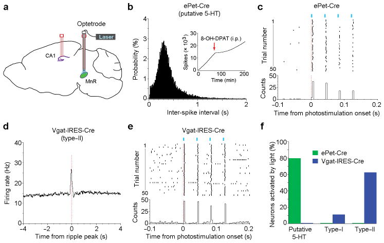
Figure 6. Optical tagging of the MnR serotonergic and GABAergic neurons.
(a), Schematic drawing showing a 4-tetrode probe in the hippocampal CA1 and a MnR optetrode consisting of an optic fiber and 4 tetrodes. ePet-Cre (n = 4) and Vgat-IRES-Cre (n = 3) mice received these implants and an injection of AAV-DIO-ChR2-EYFP into the MnR for the identification of serotonergic and GABAergic neurons, respectively. (b), Inter-spike interval histogram of a putative serotonergic neuron identified by optogenetic stimulation. Inserted panel (b) shows cumulative spikes of the same neuron after an injection of the serotonin 1A receptor agonist 8-OH-DPAT (0.2 mg/kg, i.p.). (d), Peri-ripple event histogram of a putative GABAergic neuron identified by optogenetic stimulation. (c,e), Peri-event rasters and histogram of the same 2 neurons (as shown in b and d) upon 4-pulse light trains (25 Hz; pulse width, 3 ms). (f), Percentages of putative 5-HT (n = 5 and 8 from ePet-Cre and Vgat-IRES-Cre mice, respectively), type-I (n = 6 and 9, respectively) and type-II (n = 16 and 8, respectively) neurons that responded to photostimulation. Neurons are considered to be responsible to photostimulation if peri-stimulation histogram bin with the z-score of 3.28 (P < 0.001) or greater occurs within 5 ms from the onset of photostimulation. Response latencies varied between 2 and 5 ms.

