Fig. 1.
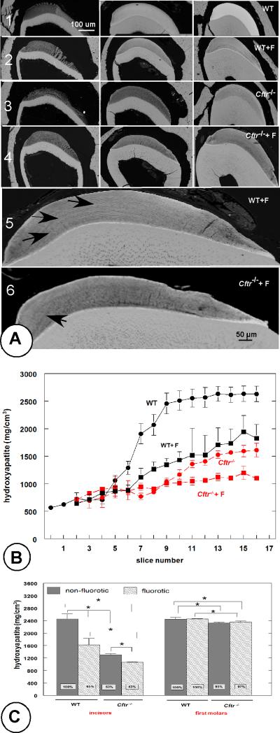
Fig 1a. Backscattered-electron detector (BSC) microscopy. Effect of Cftr-null mutation on enamel mineralization in lower incisors from fluorotic and nonfluorotic Cftr-null mice. Backscatter electron microscopy (Fig. 1a), and MicroCT (Fig. 1b, c). Fig. 1a. Row 1: nonfluorotic wild-type mouse; row 2: fluorotic wild-type mouse; row 3: non-fluorotic Cftr-null mouse and row 4: fluorotic Cftr-null mouse. The left column (from rows 1-4) represents secretion stages, the central column mid-maturation and the right column late maturation stages. Row 5 (fluorotic wild-type mouse) and row 6 (fluorotic Cftr-null mouse) show weak incremental lines (arrows).
Fig. 1b. Microcomputed tomography. Effect of fluoride on mineral density of incisor enamel from Cftr-null and wild-type mice as function of development (300 μm intervals). Slices 1-3 are secretory stage, from slice 4 onwards: maturation stage. Filled circles represent WT (black; solid line) and Cftr-null enamel (red, interrupted line). Filled squares (black, solid line): fluorotic WT and fluorotic Cftr-null enamel (red, interrupted line) (n=3). All density values of non-fluorotic Cftr-null and fluorotic wild-type enamel after slice 7 are significantly lower than nonfluorotic wild- type enamel.
Fig. 1c. Microcomputed tomography. Mineral density of incisor and first molar enamel from Cftr-null mice. Molar enamel is barely affected in contrast to the incisor enamel from the same animals. Fluoride exposure started when enamel formation in first molars was completed. Averages and standard deviation (slices 10-16; 3 mice/ group).* p < 0.05.

