
Figure 1.
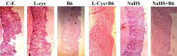
Histological evaluation of gastric mucosa. Representative gastric sections were obtained 1 hr after ethanol administration. C-E: Control group indicate severe disruption to the upper half of mucosal thickness and necrotic lesions penetrating deeply into mucosa; L-Cys, B6, NaHS, L-Cys+B6, NaHS+B6: animals pretreated with L-cysteine (100 mg/kg, IP), vitamin B6 (10 mg/kg, IP), NaHS (80 µg/kg, IP), L-cysteine (100 mg/kg, IP)+vitamin B6 and NaHS (80 µg/kg, IP) +vitamin B6, demonstrate moderate disruption of the surface epithelium. All of the sections stained with hematoxylin and eosin; ×100 magnification