
Figure 3.
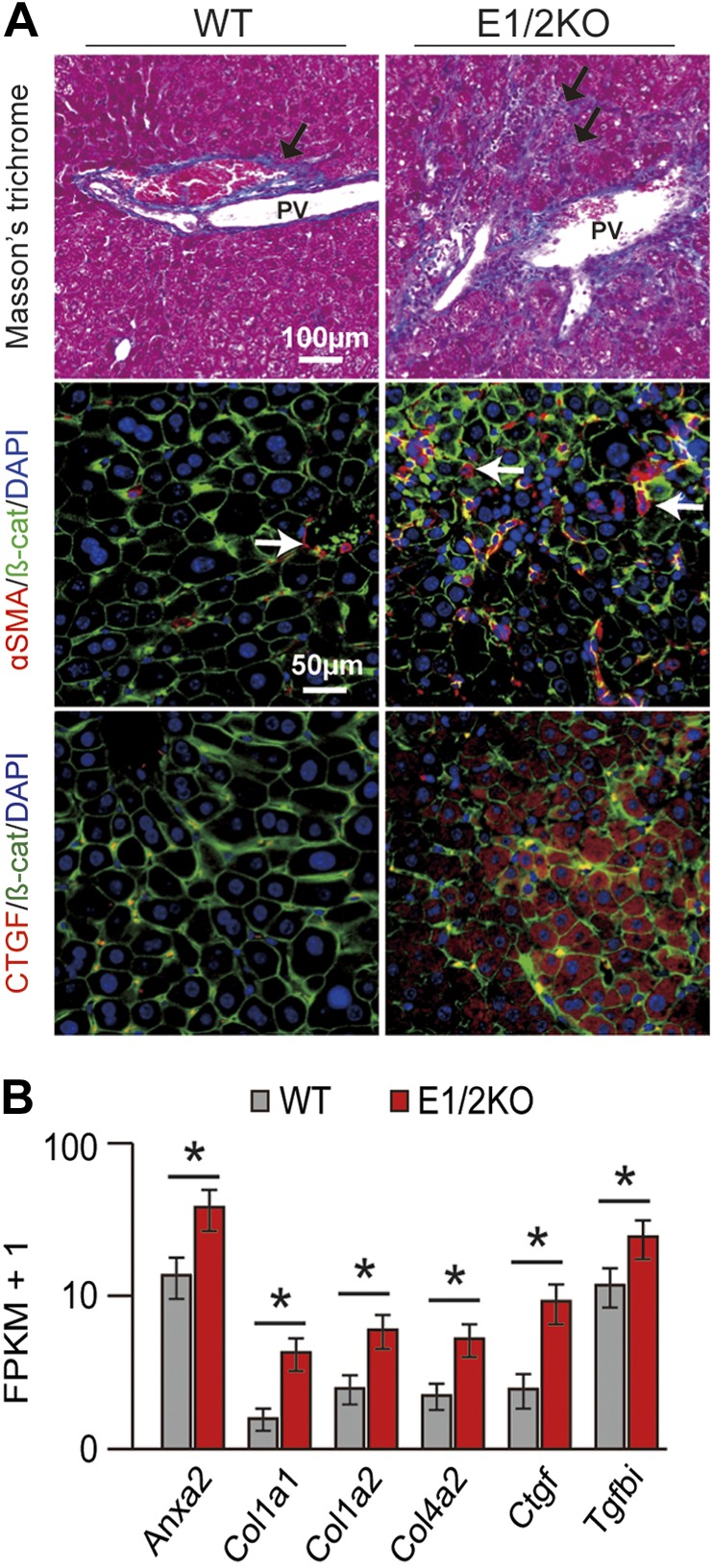
E1/2KO mice developed liver fibrosis by 8 mo of age. A) Top panels show Masson’s trichrome staining on paraffin-embedded liver sections revealing increased density of collagen fibers (arrows) in portal and periportal areas in E1/2KO livers. PV, portal vein. Middle and bottom panels show representative double-immunofluorescence staining for stellate cell marker, αSMA (red) and fibrogenic growth factor, CTGF (red) with β-catenin (β-cat; green). Nuclei were counterstained with DAPI. B) Up-regulation of fibrosis-related genes in E1/2KO livers as determined by RNA-seq. Cuffdiff (14) was used to detect significantly up-regulated genes with an FDR-adjusted P value of 0.05 (asterisk).