
Figure 6.
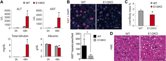
The genetic loss of EZH1 and EZH2 increases hepatocyte damage and reduces hepatocyte proliferation during compensatory liver regeneration induced by two-thirds PHx. A) Serum levels of ALT, AST, and total bilirubin are increased and levels of albumin decreased in E1/2KO mice at 48 h after PHx. B) Top panels show representative immunofluorescence staining for cell proliferation marker Ki67 (red). Nuclei were counterstained with DAPI. Bottom panels show quantification of Ki67-positive hepatocytes. The number of Ki67-positive cells was determined in 5 independent confocal images taken at ×200 magnification and expressed as numbers per field. Data are the mean ± sd (n = 5 mice for each genotype). *P < 0.05 as compared to the age-matched WT. C) Reduced recovery of liver mass in E1/2KO mice calculated as percentage of body mass. D) Representative H&E staining. Arrows indicate dividing hepatocytes. Scale bars in (B) and (D) are 50 and 100 μm, respectively.