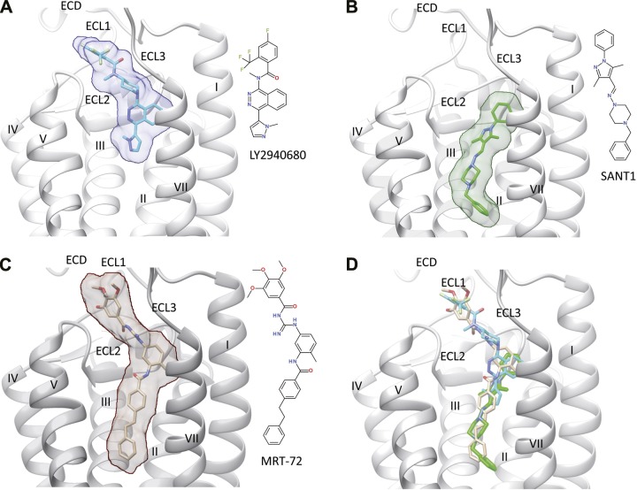
Figure 1.
Binding of different antagonists (A, LY2940680; B, SANT-1; C, MRT-92) to the transmembrane domain of hSmo (white ribbons). The ECD, the 3 extracellular loops (ECL1, ECL2, ECL3), and the 7 transmembrane helices (I–VII) are labeled, with the exception of helix VI, which is masked for the sake of clarity. The bound ligand is indicated by sticks and rendered by a transparent surface. The inset illustrates the structure of each ligand. D) All 3 ligands are simultaneously rendered in the binding site. A, B) X-ray structures (PDB accession codes 4JKV and 4N4W, respectively) (2, 3). C) Obtained by docking MRT-92 into the SANT-1-bound hSmo structure.

