
Figure 4.
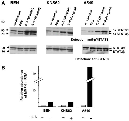
IL-6–inducible tyrosine phosphorylation of STAT3 and concomitant transcriptional activation of the MMP-1 gene in lung cancer cell lines. (A) Cell lines BEN, KNS62, and A549 were grown to 90% confluency in full medium, then starved from FCS for 6 hours, and subsequently treated optionally for 30 minutes with 10% FCS or different concentrations of IL-6 as indicated. Cells were then lysed and extracts were separated, blotted, and analyzed for STAT3 phosphorylation and expression as in Figure 3. (B) IL-6–dependent MMP-1 expression in A549 lung cancer carcinoma cells. Cells were grown for 24 hours in the absence or presence of 20 ng/ml IL-6 as indicated. Total RNA was isolated, reverse transcribed into cDNA, and quantified by reverse transcription–PCR with specific MMP-1 primers and probes. Signal intensities derived from respective non-stimulated cells were set to 1. Results from a typical experiment out of at least three independent experiments are shown.