
Figure 1.6.
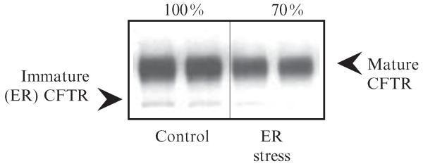
CFTR detection by Western blot. Representative results of CFTR protein detection in whole cell lysates by Western blot. Total cell lysates (15 μg) were separated by 8% PAGE, Western transferred and CFTR was detected using MM13-4 anti-CFTR monoclonal antibody, anti-mouse IgG-HRP (Pierce), and ECL (Pierce). Samples were analyzed in duplicates. Densitometry (Scion Image) was performed to assess CFTR expression levels in control and ER-stressed Calu-3 cells (immature CFTR, the core glycosylated ER form of CFTR; mature CFTR, the fully glycosylated, post-ER form of CFTR).