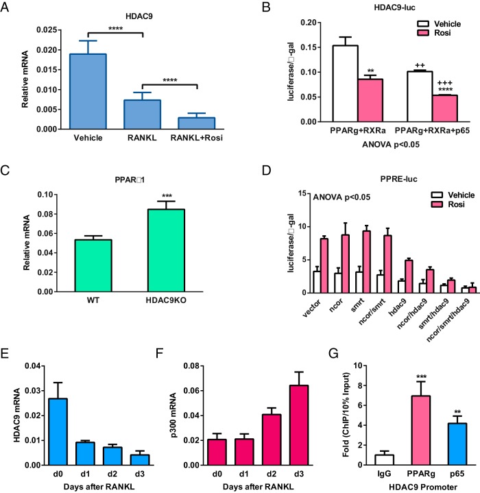
Figure 4.
Reciprocal suppression between HDAC9 and PPARγ/RANKL signaling. A and B, PPARγ activation and RANKL signaling inhibit HDAC9 expression. A, HDAC9 expression was decreased by RANKL and further down-regulated by rosiglitazone (Rosi) in osteoclast differentiation cultures (n = 5). B, Readout from a luciferase reporter driven by a 921-bp HDAC9 promoter fragment was reduced by Rosi and further diminished by the NFκB subunit p65. HEK293 cells were transfected with PPARγ, RXRα, and/or p65, together with CMV-β-gal as an internal control, and then treated with Rosi or vehicle control next day; luciferase activity was quantified 24 hours later and normalized by β-gal activity (n = 6). C, HDAC9 inhibits PPARγ1 expression. PPARγ1 expression was elevated in HDAC9-KO bone marrow osteoclast differentiation cultures compared with WT control cultures (n = 5). D, HDAC9 inhibits PPARγ activity together with corepressors SMRT and NCoR. CV-1 cells were cotransfected with PPRE-luc reporter and expression vectors for PPARγ, RXRα, HDAC9, NCoR, and/or SMRT or vector control, as well as a CMV-β-gal reporter as internal control (n = 6). E and F, During RANKL-induced osteoclast differentiation, a decrease in HDAC9 expression (E) was accompanied by an increase in the expression of a HAT p300 (F) (n = 5). G, Chromatin immunoprecipitation (ChIP) analysis of the binding of PPARγ and p65 to HDAC9 promoter in bone marrow osteoclast differentiation cultures on day 6 (n = 4). Error bars, SD.

