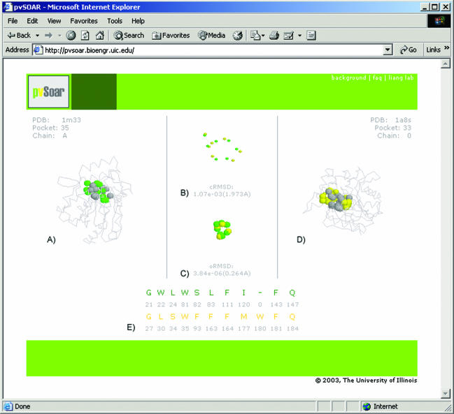
Figure 1.
Interactive environment for the visualization of pvSOAR search results. This example depicts a pocket surface pattern of (A) protein BioH from E.coli (CASTp pocket id = 35, PDB id = 1m33) and (D) haloperoxidase (CASTp pocket id = 33, PDB id = 1a8s) from Pseudomonas fluorescens. The superposition of conserved pocket residues and the superposition after projection on to a unit sphere are shown in (B) and (C), respectively. The alignment of sequence patterns of the pocket residues is shown in (E). These two surface patterns share strong similarity (cRMSD P-value = 1.073 × 10⁻³, oRMSD P-value = 3.836 × 10⁻⁶), indicating that there may be functional similarity between BioH protein and haloperoxidase.

