Figure 6.

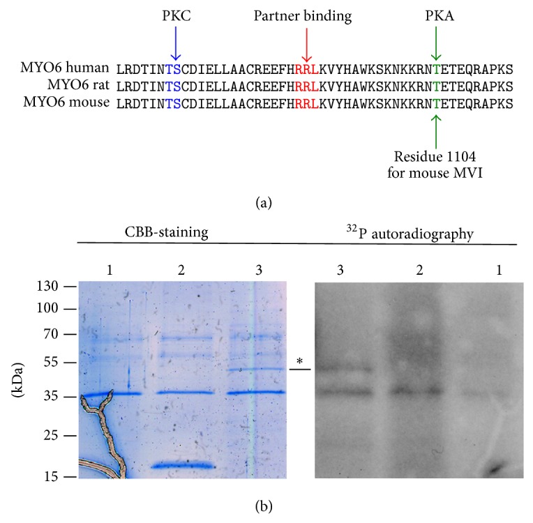
Myosin VI is a target of PKA. (a) A putative PKA phosphorylation site (threonine residue 1104 in the mouse MVI heavy chain, in green) is located in the vicinity of a very conserved region within the RRL partner recognition cluster (in red). In blue, the putative PKC phosphorylation sites. The kinase specific phosphorylation sites were depicted with a NetPhosK 1.0 Server (http://www.cbs.dtu.dk/services/NetPhosK/). The MVI sequences used for analyses are human (Q9UM54), rat (D4A5I9), and mouse (Q64331). (b) In vitro PKA phosphorylation assay. Left panel: Coomassie brilliant blue stained gel (CBB-staining), right panel: 32P autoradiogram. Lane 1: the catalytic kinase domain; lane 2: GST; lane 3: GST-tagged MVI globular tail domain. ∗ Points to the ~52-kDa band of interest (GST-MVI domain).
