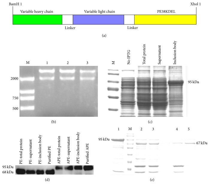
Figure 2.
Construction, expression, and purification of immunotoxin. (a) Schematic representation of immunotoxin. (b) Restriction enzyme analysis of the expression plasmid (pGEX-4T1-scFv2A9-PE). Plasmids (lanes: 1–3) were digested with BamH-I and Xhol-I, and the 2,000 bp fragment was the recombinant immunotoxin. Lane: (M) LD2000 DNA ladder. (c) SDS-PAGE of recombinant protein. Protein from noninduced cells, IPTG induced cells, and supernatant and inclusion bodies were separated on 10% agarose gel and stained with Coomassie Brilliant Blue. Lane: (M) protein marker. (d) The recombinant protein was tested via Western blot. APE represents the immunotoxin; PE represents the empty control (pGEX-4T1-PE). (e) The purified recombinant protein was digested with thrombin. After removing the GST tag and thrombin, the immunotoxin was confirmed to be 67 kDa. Lanes: (1) purified APE protein; (M) protein marker; (2) APE after digestion with thrombin; (3) APE after digestion without thrombin; (4) APE without thrombin and GST protein; and (5) purified APE without the GST tag.

