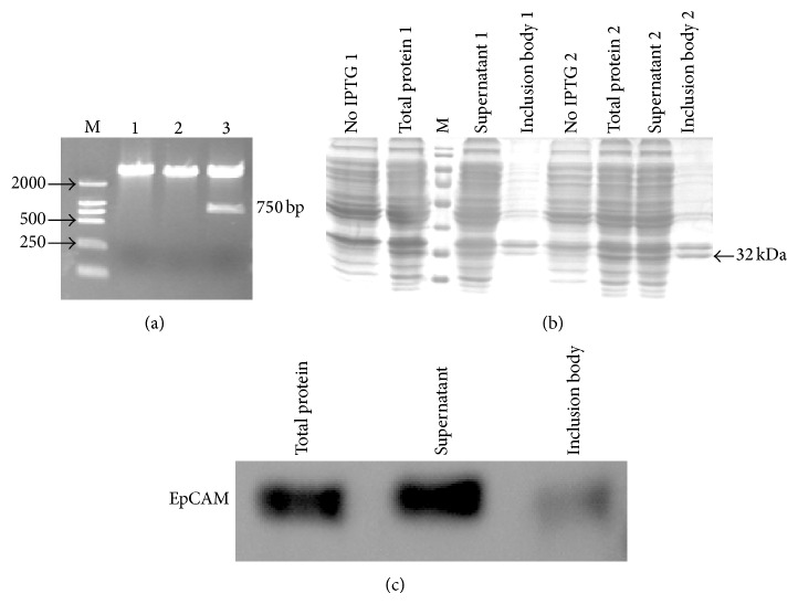
Figure 4.
Restriction enzyme analysis, SDS-PAGE, and Western blot of recombinant EpCAM expressed in M15 E. coli cells. (a) The plasmid pQE30-EpCAM was digested with Kpn I and Hind III. Lanes: (M) LD2000 DNA marker; (1–3) digestion products 1 and 2 were negative colonies; 3 was the positive colony. (b) Proteins were separated by 10% SDS-PAGE and visualized by Coomassie Brilliant Blue R250 staining. Two colonies were induced to express protein. (c) Purified HIS-EpCAM was identified by Western blot.

