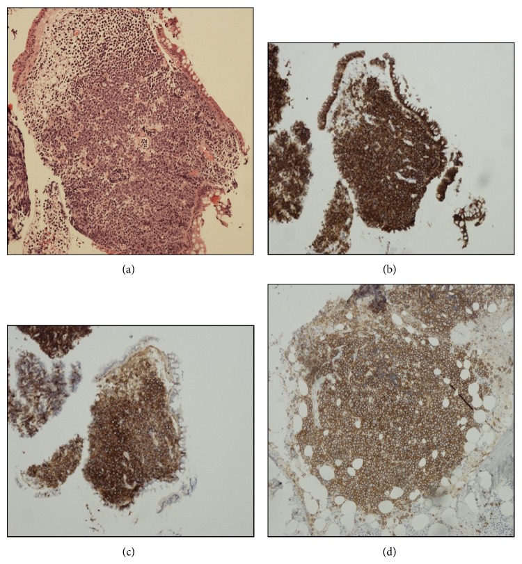
Figure 3.
Histological assessment of the colonic mass and bone marrow trephine; (a) H&E ×10 showing diffuse infiltration of the colonic mucosa by large blastic cells with expression of CD138 (×4) (b) and CD56 (×4) (c). Focal infiltration by CD138 (×10) expressing blastic cells in the bone marrow trephine biopsy (d).

