Abstract
The poly(A) tail is found on the 3’-end of most eukaryotic mRNAs, and its length significantly contributes to the mRNAs half-life and translational competence. Circadian regulation of poly(A) tail length is a powerful mechanism to confer rhythmicity in gene expression post-transcriptionally, and provides a means to regulate protein levels independent of rhythmic transcription in the nucleus. Therefore, analysis of circadian poly(A) tail length regulation is important for a complete understanding of rhythmic physiology, since rhythmically expressed proteins are the ultimate mediators of rhythmic function. Nevertheless, it has previously been challenging to measure changes in poly(A) tail length, especially at a global level, due to technical constraints. However, new methodology based on differential fractionation of mRNAs based on the length of their tails has recently been developed. In this chapter, we will describe these methods as used for examining the circadian regulation of poly(A) tail length and will provide detailed experimental procedures to measure poly(A) tail length both at a the single mRNA level and the global level. Although this chapter concentrates on methods we used for analyzing poly(A) tail length in the mammalian circadian system, the methods described here can be applicable to any organisms and any biological processes.
Keywords: Circadian, Poly(A) tail length, Post-transcriptional, Oligo(dT) chromatography, Poly(A) tail (PAT) assay, LM-PAT, Poly(A)denylome
Introduction
Post-transcriptional gene regulatory mechanisms allow modification of gene expression after transcripts are made from DNA, and this type of regulation gives tremendous flexibility in overall gene expression including when, where and how much protein product is generated. Post-transcriptional processes include many different mechanisms, such as capping, splicing, 3’-end cleavage and polyadenylation, localization, translation, and ultimate turnover of the mRNA, and circadian clocks have been shown to extensively utilize post-transcriptional regulation for rhythmic regulation of gene expression, influencing many of these steps (Kojima, Shingle, & Green, 2011). The changes in poly(A) tail length of mRNAs is one of the important post-transcriptional regulatory steps that the circadian clock uses to control rhythmic gene expression. Poly(A) tails at the 3’ends are hallmarks of most eukaryotic mRNAs, and are implicated in many aspects of mRNA function, such as mRNA stability and translation efficiency. Changes in poly(A) tail length can occur throughout the lifetime of an mRNA both in the nucleus and the cytoplasm, and the balance between deadenylation and polyadenylation ultimately determines the poly(A) tail length (Eckmann, Rammelt, & Wahle, 2011). Dynamic variation in poly(A) tail length is a powerful mechanism for driving rhythmic protein expression, and therefore developing sensitive assays that can monitor changes in poly(A) tail length is important.
To date, accurate measurement of poly(A) tail length has been technically challenging, especially at the genome-wide level, due to the homogenous nature of poly(A) tails. To overcome this issue, we developed a novel genome-wide method called “Poly(A)denylome” analysis to measure changes in poly(A) tail lengths of individual mRNAs in an unbiased manner, and to identify mRNAs that have diurnal rhythmicity in their poly(A) tail length (Kojima, Sher-Chen, & Green, 2012). Using this technique, we discovered that approximately 2.5% of mRNAs in mouse liver have rhythmic poly(A) tail lengths, thus providing evidence that the circadian clock globally regulates this post-transcriptional modification. Most importantly, we also demonstrated that the fluctuation in the poly(A) tail length can ultimately drive the rhythms in the amount of protein produced (Kojima et al., 2012).
Measurement of poly(A) tail length at a genome-wide level
Since the emergence of microarrays, genomic approaches have been widely utilized to examine expression of tens of thousands of genes simultaneously. Even though the primary focus of developing these tools was to analyze differences in gene expression levels of two or more independent samples, innovative applications have subsequently enabled the measurement of other events such as DNA methylation, transcription factor binding sites, alternative splicing, and poly(A) tail length (Heller, 2002). For example, a method we developed, called “Poly(A)denylome” analysis, can identify mRNAs that have different poly(A) tail lengths, and has successfully detected mRNAs that undergo rhythmic changes in their poly(A) tail length around the circadian clock.
Poly(A)denylome analysis consists of three different components; RNA fractionation according to poly(A) tail size, 3’-end label assay to validate the fractionation, and microarray analysis. First, total RNAs are selected by the presence of poly(A) stretches, and then these RNAs are divided into two different populations: one has short poly(A) tail lengths of approximately ~70nt or less, and the other has poly(A) tail lengths that are longer than ~60nt. This length threshold can be modified by changing the salt concentration in the elution buffer (see below for details). The poly(A) tail length in each population as well as the RNA integrity need to be further verified by the 3’-end label assay. Following the validation and clean-up of these RNAs, they can be subjected to microarray analysis. Needless to say, microarrays are tools to quantify mRNA levels, not to obtain poly(A) tail length information. Therefore, relative poly(A) tail length information is derived from the ratio of the level of each mRNA in the long poly(A) tail length population over the level in the short poly(A) tail length population. We have shown that these ratios accurately reflect the dynamics of poly(A) tail length changes and have successfully used these ratios to define whether a specific transcript has a rhythmic poly(A) tail length, based on certain threshold criteria, such as basal expression level, a fit to a sinusoid, or amplitude of rhythms (Kojima et al., 2012).
In this chapter, we will describe detailed procedures for the RNA fractionation and 3’-end labeling assay below. However, we will not cover the detailed techniques for microarray analyses here, as there are well documented standard protocols, although we will provide some information specific to Poly(A)denylome analysis.
Poly(A) tail size RNA fractionation
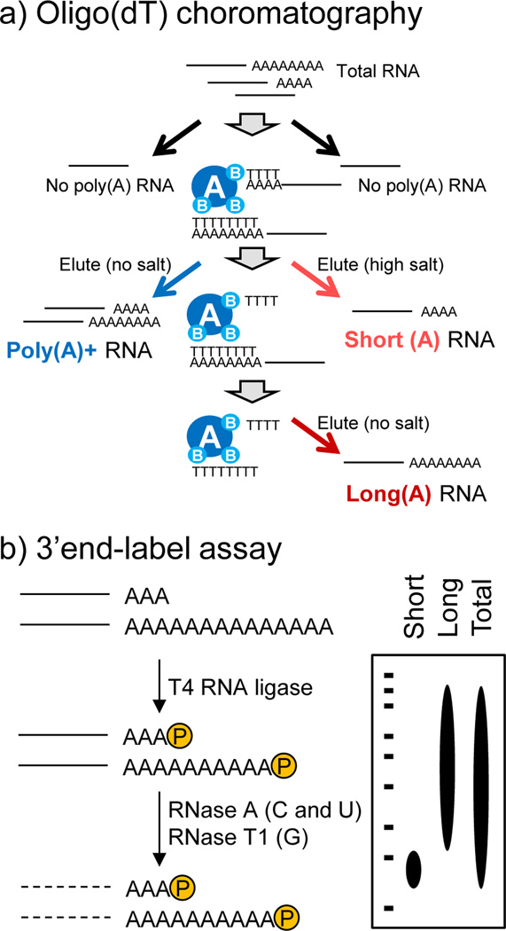
The first step is to employ poly(A) tail size RNA fractionation, which divides RNAs into populations that have either short or long poly(A) tail length. This is done by oligo(dT) chromatography where oligo(dT)-bound poly(A)+ RNAs can be differentially recovered using elution buffers with different salt concentrations, owing to the difference in affinity of long and short tails for the oligo(dT) beads, as reported previously (Meijer et al., 2007). Another similar method for poly(A) tail length fractionation that utilizes the temperature, instead of salt concentration, has also been developed (Beilharz & Preiss, 2009), however, we found that varying the salt concentration is easier and more reproducible than changing the temperature, partly because ambient temperature can be slightly different each day and hard to strictly control. It is also important to keep in mind that all the reagents must be prepared as RNA-grade (i.e. DEPC-treated), as the success of RNA fractionation depends on the integrity of the RNAs. This is particularly important for the RNA fractionation step, since the elution step needs to be performed at room temperature (20–25°C) overnight. RNA can be extracted by any conventional methods, but a significant amount of starting materials (we used 80 ug) are needed to be able to visualize the bulk poly(A) tail length at a later step (see also 3’-end label assay). As a general guideline, 100 mg of mouse liver tissue with 1 mL TRIZOL (Life Technologies) yields 300–400 ug of RNA.
- Prepare dilution buffer by mixing the following and preheat at 70°C.
300 ul 20×SSC 10 ul 1M Tris-HCl (pH 7.4) 2 ul 0.5M EDTA 25 ul 10% SDS 10 ul β-Mercaptoethanol 653 ul DEPC-DDW Mix 80 ug RNA sample (in a volume of 40 ul or less) with 400 ul GTC buffer, 8 ul β-Mercaptoethanol, 15 ul biotinylated oligo(dT) (50 pmol/ul), and 816 ul Dilution buffer (prepared in step 1).
Incubate the sample mixture at 70°C for 10 min.
Spin at 12,000 g for 10 min at room temperature. Note that there might be white precipitant after the spin, which should be avoided. If there is a white crystallized material floating instead, tap these and spin down again to remove. Collect the supernatant.
Meanwhile, pre-wash the magnetic beads with 600 ul 0.5×SSC 3 times, and keep the last 0.5×SSC in a tube until beads are ready for the next step.
After carefully removing the 0.5×SSC, add the supernatant from 4) to the magnetic beads. Try not to collect any of the white precipitants, as this could stick to magnetic beads and hinder the RNA-oligo(dT) interaction.
Incubate for 15 min at room temperature with gentle mixing or rotation.
Take out the supernatant and save this as an “unbound fraction” for later trouble-shooting, if necessary.
Wash the magnetic beads with 600 ul 0.5×SSC 3 times at room temperature, with at least 5 min mixing between each wash.
After the third wash, add 200 ul 0.075×SSC to the magnetic beads and incubate for 2 hrs at room temperature to elute the short tailed RNAs. After collecting the eluent, add another 200 ul 0.075×SSC and incubate with the magnetic beads overnight at room temperature for complete elution of short-tailed RNAs. (Therefore, the combined 400 ul of eluent from these two steps contains the short poly(A) tailed RNA population). Then add 400 ul DEPC-DDW and incubate for 2 hrs at room temperature to collect long poly(A) tailed RNAs. Alternatively, 400 ul DEPC-DDW can be directly added to the magnetic beads immediately after the washing step (step 9) to collect poly(A)+ enriched (total non-fractionated) RNAs.
Recovered RNAs can be further cleaned by RNeasy MinElute Cleanup Kit, if necessary. Alternatively, these RNAs can also be precipitated with a conventional method (i.e. isopropanol precipitation) in order to further purify and concentrate the RNAs.
3’-end labeling assay
This assay examines the poly(A) tail length of a bulk RNA pool. Typically, we perform this assay immediately after poly(A) size fractionation to check and validate the poly(A) tail length difference in each pool. To this end, the 3’-end of the poly(A) tail is radiolabeled followed by digestion of the body of the RNAs with both RNase A that cleaves single-stranded C and U residues and RNase T1 that cleaves G residues, leaving the poly(A) tail intact. Digested RNAs can therefore be separated on polyacrylamide gels and the poly(A) tail length can be visualized by autoradiography. For the labeling of the 3’-end of poly(A) tails, [5’-32P] pCp molecules are ligated to the RNAs. [5’-32P] pCp is commercially available, although costly. Therefore, it is highly recommended to make [5’-32P] pCp on your own by phosphorylating CMP molecules instead, which is fairly straightforward and simple, and reduces the cost significantly. Therefore, a protocol to produce [5’-32P] pCp is also provided below.
- To prepare [5’-32P] pCp, mix the following:
2.5 ul 1 M Tris-HCl (pH7.4) RNA grade 10.0 ul 50 mM MgCl2 2.5 ul 0.1 M DTT 1.0 ul 7.5 mM CMP dissolved in DEPC-DDW (pH9.0) and filtered (0.22 um) 1.0 ul 75 mM Spermine dissolved in DEPC-DDW and filtered (0.22 um) 5.0 ul [γ-32P] ATP 2.0 ul T4 polynucleotide kinase 26.0 ul DEPC-DDW to make a total volume of 50 ul Incubate the mixture at 37°C overnight and then heat-deactivate the enzyme by heating at 90°C for 3 min. This mixture can be stored at −20°C until use, however, handle it with caution as this contains radioisotope materials.
- 3’-end label RNAs by mixing the following:
5.0 ul 10× T4 RNA ligase Buffer 5.0 ul 10 mM ATP 5.0 ul DMSO 5.8 ul Glycerol (RNA grade) 10.0 ul [5’-32P] pCp (made in steps 1 and 2 above) 2.0 ul T4 RNA ligase 17.2 ul RNA with DEPC-DDW to make a total volume of 50 ul Incubate this labeling mixture at 4°C overnight. Note that this contains radioactive materials, so the incubation needs to be performed carefully.
- Digest RNA bodies by incubating the following for 30 min at 37°C:
40.0 ul labeling mixture (from section 3 and 4 above, save the other 10 ul for step 7) 12.0 ul 5 M NaCl 2.0 ul 0.5 M EDTA 2.0 ul RNase A/T1 mixture 1.0 ul yeast tRNA (10 mg/ml) 143.0 ul DEPC-DDW to make a total volume of 200 ul Incubate the mixture at 37°C for 30 min.
Add 190 ul of DEPC-DDW to the 10 ul RNA leftover from 3’end labeling. These will serve as a control for the labeling and should be run on the gel with RNase digested samples (step 10).
Both digested and undigested RNAs will be extracted by TRIZOL, precipitated by isopropanol, and dissolved in 5 ul DEPC-DDW using a standard procedure.
Mix RNA samples with 2× gel loading buffer.
Heat the samples at 95°C for 5 min then keep on ice.
Run samples on 7.5% denaturing mini-gels (see recipe in “Materials”) until the dyes run off. Generally, it takes approximately 1 hour to finish when the voltage is set at 120V.
Microarray analysis
It is crucially important to choose the right microarray platform that does not require poly(A)+ selection during the RNA/cDNA preparation process when performing Poly(A)denylome analysis, as each RNA pool (short vs long vs non-fractionated) has different poly(A) tail lengths and differences in poly(A) tail length could introduce a bias during sample preparation. In addition to this technical concern, the analytical method is also critical in determining whether a given transcript exhibits the poly(A) rhythmicity. The status of relative poly(A) tail lengths can be obtained as a ratio of gene expression level in the long poly(A) pool over the short poly(A) pool. Therefore, the circadian fluctuation of the ratio needs to be calculated to detect whether a particular transcript has a rhythmic pattern. Presence of rhythmic patterns can be determined by several algorithms such as JTK_CYCLE, ARSER, COSOPT, Fisher’s G test, and CircWave (http://www.euclock.org/results/item/circ-wave.html) (Hughes, Hogenesch, & Kornacker, 2010; Panda et al., 2002; Yang & Su, 2010), although each algorithm has unique characteristics, and multiple factors such as tolerance to noise (i.e. outliers), and a fit to a sinusoid can significantly affect the results. This is particularly important for Poly(A)denylome analysis, because the long/short ratio, a poly(A) tail length indicator, is far less robust in rhythmicity and much more noisy compared to circadian fluctuation of mRNA expression. Therefore, it is highly recommended to use much less stringent parameter settings compared to conventional transcriptome analyses, in order to obtain poly(A) rhythmic genes. Any false positives can be removed by independent validation of the tail length rhythms using methods described below.
Measurement of poly(A) tail length at a single-gene level
Poly(A) tail (PAT) assay
Compared to large-scale analyses, measuring the poly(A) tail length at the single mRNA level has been much more well developed, and a variety of methods, generally called Poly(A) tail (PAT) assays, are currently available. A classic example, Oligo(dT)/RNase H Northern, compares the mRNAs with poly(A) tails against those devoid of poly(A) tails using oligo(dT) and RNase H, as the oligo(dT) hybridizes to the poly(A) tail of the mRNAs and RNase H then recognizes and cleaves the RNA:DNA hybrids (Salles & Strickland, 1999). A second antisense oligodeoxynucleotide that targets the sequence near the 3’end of the RNA can be also included in the reaction for better visualization of the changes in the poly(A) tail by cleaving off the majority of RNA body. This method, however, is labor-intensive and requires a large amount of RNA. In addition, it is difficult to detect small changes in poly(A) tail length, especially when your gene of interest has a long transcript. More recent techniques are all based on RT-PCR, rather than Northern Blot, and successfully overcome these issues by providing increased sensitivity, speed and length quantification. Several methods, such as RACE-PAT (Salles, Darrow, O'Connell, & Strickland, 1992), and Ligation-Mediated (LM)-PAT (Salles & Strickland, 1995), extension PAT (ePAT) (Janicke, Vancuylenberg, Boag, Traven, & Beilharz, 2012) or splint-mediated PAT (sPAT) (Minasaki, Rudel, & Eckmann, 2014), have been described, and these are slightly different in their technology in the cDNA synthesis step, each having advantages and disadvantages.
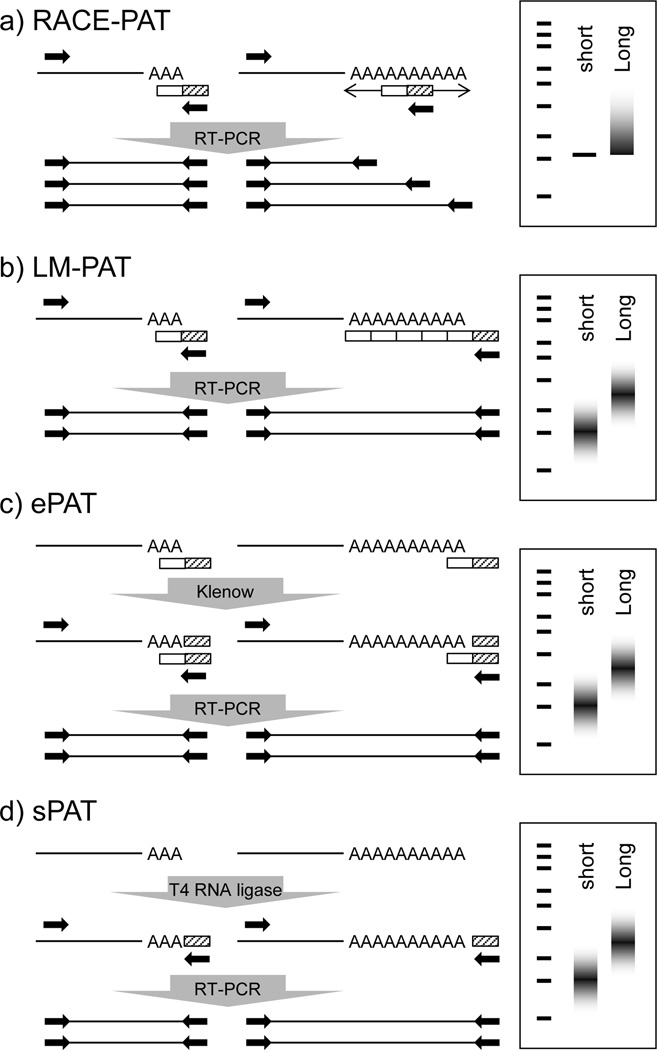
The RACE-PAT assay derives from the 3’RACE (rapid amplification of cDNA ends) protocol, where RNA is reverse-transcribed with an oligo(dT) primer linked to a G/C-rich anchor sequence. Since hybridization of the oligo(dT) can occur anywhere along the entire length of the poly(A) tail, a heterogeneous pool of cDNAs primed at all possible positions along the poly(A) tail will be synthesized (Figure 2a). In contrast, LM-PAT targets the oligo(dT) anchor to the 3’-end of the poly(A) tail by saturating the poly(A) tail being annealed with phosphorylated oligo(dT)12–18 [p(dT)12–18], which are ligated together in the presence of T4 DNA ligase (Figure 2b). Potential problems with these two methods include the possibility of internal priming of the oligo(dT) anchor primer to internal A-rich stretches during cDNA synthesis and/or transcripts arising from alternative polyadenylation site usage within the same gene. Theoretically however, PCR products derived from internal poly(A) stretch priming or alternative polyadenylation should be easily distinguishable from that of the poly(A) 3’-end priming products, as these would result in bands of different size. More recent methods, ePAT and sPAT, were developed to avoid this internal priming issue. With ePAT, the oligo(dT) anchor is first annealed to poly(A) tails at a high temperature (55°C), ensuring the anchor can only stay annealed at the end of the poly(A) tails. Klenow polymerase and dNTPs are then added to the reaction, resulting in template extension of the 3’-end of the poly(A) tract (Figure 2c). Finally, sPAT utilizes an RNA (not DNA) anchor sequence ligated immediately after the 3’-end of the poly(A) tail using a single-strand DNA splint as a bridge (Figure 2d), thus avoiding the internal priming while targeting the anchor to the end of poly(A) tail. Subsequent PCR amplification of this cDNA pool with a message-specific primer and the oligo(dT) anchor yields a mixture of products representing a variety of different poly(A) tail lengths of the specific transcript of your interest.
Figure 2. Schematic representations of various PAT assays and their ideal results.
a) RACE-PAT. Due to the ability of the oligo(dT) anchor to prime reverse transcription from anywhere along the poly(A) tail, the results appear as a solid smear due to annealing from the 5’-most start of the poly(A) tail to the 3’-end. While b) LM-PAT, c) ePAT, and d) sPAT can easily identify discrete changes in poly(A) tail length over time, due to targeting of the oligo(dT) anchor to the 3’- end by oligo(dT) ligation. The half-white/half-diagonally hatched box represents the oligo(dT) anchor for reverse transcription, the open box (in LM-PAT) represents oligo p(dT)12–18 subunits, the hatched box (in sPAT) represents an RNA anchor, and the closed box represents the message-specific primer for PCR.
Message-specific primers should be suitable for any types of PAT assays, although there are basic criteria that need to be met as this is the only parameter that dictates the target specificity. Ideally, primers should be designed 250–500 nt upstream of the 3’-end of the mRNA (i.e. poly(A) signal), and include a unique restriction site (within the amplified region) approximately 50–200 bp downstream from the message-specific primer to test amplification specificity. It is better to design a primer that yields a single shorter band representing the 5’- fragments and a slower-migrating heterogeneous 3’-end smear, but it is also tolerable to have longer 5’-fragments as long as they do not overlap with the poly(A) tail smear derived from the 3’-fragment, the size of which is sometimes hard to predict. Depending on the expression level and sequence near the 3’-end of your mRNA of interest, it might take several different attempts to find primers that confer enough specificity to give rise to a single band. However, it is also possible to have two or more specific bands derived from alternative polyadenylation, which occurs in more than 50% of human genes (Tian, Hu, Zhang, & Lutz, 2005). It is also worth noting that the restriction enzyme sites might be altered due to SNPs, therefore, it is encouraged to try multiple different enzymes, if possible, especially when the message-specific primer yields a single band.
Standard PCR conditions should work for most PAT assays. The annealing temperature and cycle numbers must be empirically determined, depending on the primer sequence and your mRNA of interest. Once the PCR products are obtained, proper amplification of the mRNA of interest can be confirmed by either restriction digest analysis as described above or direct sequencing of the PCR products. As an initial approach, electrophoretic analysis can be conducted using ethidium bromide-stained 2–3% agarose gels, which provided enough resolution and sensitivity in our study. If increased resolution and sensitivity are desired, the addition of a small amount of radiolabeled dNTP is also an option in each PCR, in which case the samples should be cleaned to remove radiolabeled dNTPs.
Potential issues with PAT assays
The biggest issue common to all PAT assays is that the poly(A) tail length measurements can be significantly affected by a difference in gene expression level between each samples, and it is extremely difficult to compare the poly(A) tail length of transcripts from two samples when their RNA expression levels are considerably different. Considering PAT assays are qualitative, rather than quantitative, it might be preferable to adjust the amount of RNAs prior to PAT assay, so that amplification efficiency and visibility on the gels may become similar.
Another thing to keep in mind is that PCR amplification favors the generation of smaller amplicons; in other words, it is biased toward shorter poly(A) tails and this is partly because DNA polymerases (i.e. Taq polymerase) generally work less efficiently in amplifying homogenous nucleotides, such as poly(A) stretches. This is particularly relevant for transcripts with long poly(A) tails but low in abundance, needing a higher number of amplification cycles. RACE-PAT, ePAT and sPAT are much more sensitive to low-abundance mRNAs, compared to LM-PAT that requires the ligation process which is not highly efficient and reduces the size of the cDNA population. However, LM-PAT, ePAT and sPAT all assure the targeting of oligo(dT) anchor to the 3’-end of poly(A) tails, regardless of the length of poly(A) tails, thus avoiding any bias towards shorter poly(A) tails.
The main technical challenge with PAT assays is achieving good PCR products. It is not uncommon to get very weak/no amplification, or high background noise, given that the message-specific primer is the only parameter that dictates the PCR specificity (as the other primer, the anchor primer, needs to be common to all target mRNAs). This problem can be sometimes be simply solved by changing PCR conditions (i.e. annealing temperature, primer concentration, a number of cycles, etc), but it often requires trying several different primers with various PCR conditions to get an optimal result. For this reason, it is highly recommended to try a test PCR using a control cDNA to validate primers and restriction enzymes, before testing the experimental samples.
LM-PAT assay
In this review, we will provide a detailed protocol for the LM-PAT assay below, as we find this technique to be most reliable and reproducible, compared to other PCR-based PAT assays. Readers interested in other methods may want to refer elsewhere (Janicke et al., 2012; Minasaki et al., 2014; Salles et al., 1992). For LM-PAT, it is highly recommended to start with poly(A)+ enriched RNAs, as this not only eliminates genomic DNA contamination, but also significantly increases the sensitivity and target specificity of the assay. Alternatively, it is also possible to use radioisotopes to increase the sensitivity.
- Pre-warm a cDNA synthesis mixture of the following at 42°C:
4 ul 5×1st strand buffer 1ul RNasin (20 U/ul) 2 ul 0.1 M DTT 1ul 10 mM dNTP 1ul 10 mM ATP 1ul T4 DNA ligase (10 U) 3 ul DEPC-DDW Mix poly(A)+ RNA (50 ng or more) and phosphorylated Oligo(dT)15 (20 ng) in a total volume of 10 ul in a 0.2ml tube.
Incubate RNA-Oligo(dT) mixture at 65°C for 10 min, then transfer to 42°C immediately. Note that thermal cyclers are not recommended for this step, as this takes several seconds to shift the temperature. The sudden change in temperature is essential (i.e. use two separate water baths closely placed)
Add 10 ul of the pre-warmed reaction mixture in 1) to the tube. Mix well by pipetting and incubate at 42°C for 30 min.
At the end of the incubation, while still at 42°C, add 1ul of Oligo(dT)12 anchor (5’-GCGAGCTCCGCGGCCGCGTTTTTTTTTTTT-3’) (200 ng/ul).
Quickly vortex, spin, and incubate the mixture at 12°C for 2 hr, then 42°C for 2 min. This step and hereafter can be done in a thermal cycler.
While still at 42°C, add 1ul of SuperScript II Reverse Transcriptase (Life Technologies), and mix well.
Incubate 42°C for 1hr and inactivate enzymes by heating at 65°C for 20 min.
- Perform a PCR as follows. The PAT first-stranded cDNAs can be diluted 1:50–1:200, for analysis of multiple mRNAs. Alternatively, these cDNAs can also be used undiluted, especially when the expression level of target mRNAs is low.
2.0 ul 10× PCR buffer 0.8 ul 50 mM MgCl2 0.4 ul 10 mM dNTP 0.4 ul Oligo(dT)anchor (25 pmol/ul) 0.4 ul Message-specific primer (25 pmol/ul) 0.2 ul Taq polymerase 1.6 ul PAT cDNA (original or diluted) 14.2 ul DDW to make a total volume of 20 ul - Recommended PCR cycle as a starter:
1X 93°C 5 min 30–40X 93°C 30 sec 60–65°C 30 sec 72°C 1 min 1X 72°C 7 min Save 10 ul of the PCR reaction mixture at 4°C or −20°C until use. Add 1 ul of restriction enzyme to the remaining 10 ul PCR mixture, and incubate 37°C for 60 min.
Run the PCR reactions on agarose gels with appropriate size markers. By running digested and undigested samples together, one should be able to see the shift by enzyme digestion in amplified DNA size.
Visualize the resulting DNA fragments by ethidium bromide staining.
Determine Poly(A) tail lengths using densitometry software (we used Alpha Multiphotoimager II with AlphaInnotech software). Since the majority of bands will appear as smears due to the heterogeneous poly(A) tail lengths, it is crucial to use some objective measurement of the size. We used the center of gravity of the PCR product (as determined by densitometry) to assign a poly(A) tail size in each lane.
Materials
Poly(A) tail size RNA fractionation
GTC buffer (Promega Z5531)
Biotinylated Oligo(dT) (Promega Z5261)
Magnetic beads (Promega Z5481) *Magnetic stands are available from Promega or Life Tehcnologies.
RNeasy MinElute Cleanup Kit (Qiagen Cat# 74204)
3’-end labeling assay
CMP (Sigma C1133)
Spermine (Sigma S1141)
T4 polynucleotide kinase (New England Biolabs M0201)
10 mM ATP (EPICENTRE)
T4 RNA ligase (EPICENTRE)
RNase A/T1 mixture (Fermentas: #EN0551)
yeast tRNA (Ambion #7118)
Gel loading buffer (Ambion ; #AM8546)
7.5 % denaturing gel (for two 8×10cm gels, 1.0mm) : Dissolve 7.2 g Urea in a mixture of 1.5 mL 10× TBE, 3.25 mL 30% Acrylamide/Bis, 3.25 mL DDW, 75 ul 10% APS, 15 ul TEMED
Concluding remarks
Rhythmic gene expression is controlled by a plethora of regulatory steps including both transcriptional and post-transcriptional mechanisms, and poly(A) tail length control is one of the driving forces to achieve rhythmic protein expression. In addition to circadian biology, the relevance of regulating poly(A) tail length has also been demonstrated in other biological processes such as cell cycle, oocyte maturation, cellular senescence and synaptic plasticity (Charlesworth, Meijer, & de Moor, 2013). Therefore, the Poly(A)denylome analysis method that we developed to originally understand the circadian poly(A) tail length has broader applications, and can be applied to other processes such as those described above in order to comprehensively identify which mRNAs are subject to poly(A) tail length regulation.
It should be noted that although our protocol utilizes microarrays, similar analyses can be done using recently developed RNA-seq based methods. For example, several recent reports have described new methods by which to comprehensively analyze the poly(A) tail length, such as TAIL-seq (Chang, Lim, Ha, & Kim, 2014), or Poly(A) Tail length (PAL)-seq (Subtelny, Eichhorn, Chen, Sive, & Bartel, 2014). Further studies using these new techniques in different tissues, cells, and organisms, will be interesting for examination of the global effect of poly(A) tail length regulation on circadian gene expression.
Figure 1. Method for fractionation of mRNAs with different poly(A) tail lengths (Kojima et al., 2012).
a) Oligo(dT) chromatography was used to separate total RNA into fractions with either short or long poly(A) tails by varying salt concentrations in the elution. Total PolyA+ (non-fractionated) RNAs can also be isolated as a reference. b) Validation of fractionation by a 3’-end label assay. T4 RNA ligase catalyzes the ligation of the 5' phosphate terminus of a nucleic acid donor to the 3' OH terminus of a nucleic acid acceptor, thus introduces radioisotope label at the 3’-end of poly(A) tails. 3’-end labeled mRNAs are then digested by both RNase A that recognizes cytidine and uridine residues and RNase T1 that recognizes guanidine residues, resulting in degradation of the RNA body but not poly(A) stretches. The size of these poly(A) stretches can be visualized by autoradiography after PAGE (polyacrylamide gel electrophoresis) analysis.
Acknowledgement
We thank all the Green lab members for helpful discussions and comments on the manuscript, especially Dr. Danielle Hyman. This work was supported by the US National Institutes of Health (R01GM090247, R21NS079986, and R01AG045795) to C.B.G., Tomizawa Jun-ichi & Keiko Fund of Molecular Biology Society of Japan for Young Scientist and the Brain and Behavior Research Foundation (NARSAD) to S.K.
References
- Beilharz TH, Preiss T. Transcriptome-wide measurement of mRNA polyadenylation state. Methods. 2009;48(3):294–300. doi: 10.1016/j.ymeth.2009.02.003. [DOI] [PubMed] [Google Scholar]
- Chang H, Lim J, Ha M, Kim VN. TAIL-seq: Genome-wide Determination of Poly(A) Tail Length and 3' End Modifications. Mol Cell. 2014;53(6):1044–1052. doi: 10.1016/j.molcel.2014.02.007. [DOI] [PubMed] [Google Scholar]
- Charlesworth A, Meijer HA, de Moor CH. Specificity factors in cytoplasmic polyadenylation. Wiley Interdiscip Rev RNA. 2013;4(4):437–461. doi: 10.1002/wrna.1171. [DOI] [PMC free article] [PubMed] [Google Scholar]
- Eckmann CR, Rammelt C, Wahle E. Control of poly(A) tail length. Wiley Interdiscip Rev RNA. 2011;2(3):348–361. doi: 10.1002/wrna.56. [DOI] [PubMed] [Google Scholar]
- Heller MJ. DNA microarray technology: devices, systems, and applications. Annu Rev Biomed Eng. 2002;4:129–153. doi: 10.1146/annurev.bioeng.4.020702.153438. [DOI] [PubMed] [Google Scholar]
- Hughes ME, Hogenesch JB, Kornacker K. JTK_CYCLE: an efficient nonparametric algorithm for detecting rhythmic components in genome-scale data sets. 2010 doi: 10.1177/0748730410379711. [Research Support, N.I.H., Extramural. [DOI] [PMC free article] [PubMed] [Google Scholar]
- Research Support, Non-U.S. Gov't] J Biol Rhythms. 25(5):372–380. [Google Scholar]
- Janicke A, Vancuylenberg J, Boag PR, Traven A, Beilharz TH. ePAT: a simple method to tag adenylated RNA to measure poly(A)-tail length and other 3' RACE applications. RNA. 2012;18(6):1289–1295. doi: 10.1261/rna.031898.111. [DOI] [PMC free article] [PubMed] [Google Scholar]
- Kojima S, Sher-Chen EL, Green CB. Circadian control of mRNA polyadenylation dynamics regulates rhythmic protein expression. Genes Dev. 2012;26(24):2724–2736. doi: 10.1101/gad.208306.112. [Research Support, N.I.H., Extramural]. [DOI] [PMC free article] [PubMed] [Google Scholar]
- Kojima S, Shingle DL, Green CB. Post-transcriptional control of circadian rhythms. J Cell Sci. 2011;124(Pt 3):311–320. doi: 10.1242/jcs.065771. [DOI] [PMC free article] [PubMed] [Google Scholar]
- Meijer HA, Bushell M, Hill K, Gant TW, Willis AE, Jones P, et al. A novel method for poly(A) fractionation reveals a large population of mRNAs with a short poly(A) tail in mammalian cells. Nucleic Acids Res. 2007;35(19):e132. doi: 10.1093/nar/gkm830. [DOI] [PMC free article] [PubMed] [Google Scholar]
- Minasaki R, Rudel D, Eckmann CR. Increased sensitivity and accuracy of a single-stranded DNA splint-mediated ligation assay (sPAT) reveals poly(A) tail length dynamics of developmentally regulated mRNAs. RNA Biol. 2014;11(2):111–123. doi: 10.4161/rna.27992. [DOI] [PMC free article] [PubMed] [Google Scholar]
- Panda S, Antoch MP, Miller BH, Su AI, Schook AB, Straume M, et al. Coordinated transcription of key pathways in the mouse by the circadian clock. Cell. 2002;109(3):307–320. doi: 10.1016/s0092-8674(02)00722-5. [DOI] [PubMed] [Google Scholar]
- Salles FJ, Darrow AL, O'Connell ML, Strickland S. Isolation of novel murine maternal mRNAs regulated by cytoplasmic polyadenylation. Genes Dev. 1992;6(7):1202–1212. doi: 10.1101/gad.6.7.1202. [DOI] [PubMed] [Google Scholar]
- Salles FJ, Strickland S. Rapid and sensitive analysis of mRNA polyadenylation states by PCR. PCR Methods Appl. 1995;4(6):317–321. doi: 10.1101/gr.4.6.317. [DOI] [PubMed] [Google Scholar]
- Salles FJ, Strickland S. Analysis of poly(A) tail lengths by PCR: the PAT assay. Methods Mol Biol. 1999;118:441–448. doi: 10.1385/1-59259-676-2:441. [DOI] [PubMed] [Google Scholar]
- Subtelny AO, Eichhorn SW, Chen GR, Sive H, Bartel DP. Poly(A)-tail profiling reveals an embryonic switch in translational control. Nature. 2014;508(7494):66–71. doi: 10.1038/nature13007. [DOI] [PMC free article] [PubMed] [Google Scholar]
- Tian B, Hu J, Zhang H, Lutz CS. A large-scale analysis of mRNA polyadenylation of human and mouse genes. Nucleic Acids Res. 2005;33(1):201–212. doi: 10.1093/nar/gki158. [DOI] [PMC free article] [PubMed] [Google Scholar]
- Yang R, Su Z. Analyzing circadian expression data by harmonic regression based on autoregressive spectral estimation. Bioinformatics. 2010;26(12):i168–i174. doi: 10.1093/bioinformatics/btq189. [DOI] [PMC free article] [PubMed] [Google Scholar]


