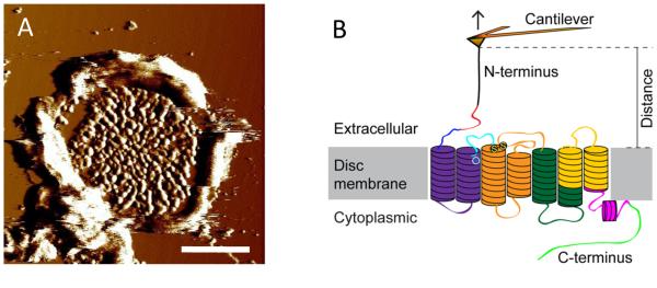
Fig. 1.

(A) An AFM deflection image of a ROS disc membrane adsorbed on mica. Scale bar, 500 nm. (B) Illustration of a SMFS experiment. The AFM probe attached to a flexible cantilever is non-specifically bound to the N-terminal end of rhodopsin embedded in ROS disc membranes. As the AFM probe is retracted from the sample surface, rhodopsin is mechanically unfolded in a stepwise manner. The stable structural segments of rhodopsin are highlighted in different colors. This figure is reproduced from (13) with permission from the American Society for Biochemistry and Molecular Biology.
