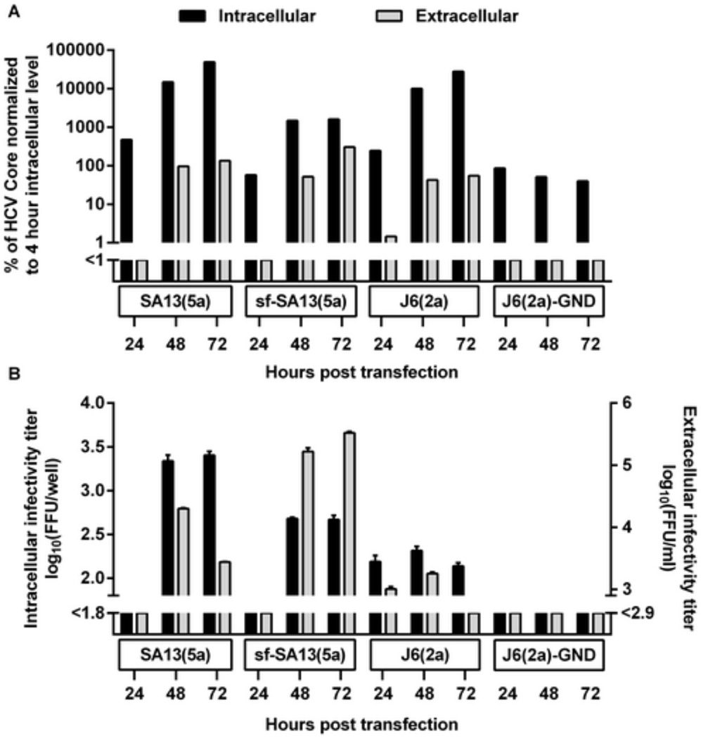
Figure 4. Serum-free culture decreased viral replication/translation but enhanced viral release and specific infectivity.
S29 cells were transfected with SA13(5a) as well as positive control (J6(2a)) and negative control (J6(2a)-GND) HCV RNA transcripts as described in Materials and Methods. (A) Intracellular (black bars) and extracellular (grey bars) Core levels were determined 24, 48 and 72 hours post transfection. Core levels were normalized to intracellular Core levels measured 4 hours post transfection. (B) Intracellular (black bars) and extracellular (grey bars) infectivity titers were determined 24, 48 and 72 hours post transfection. Infectivity titers are shown as the means (FFU/well) of three replicates with SEM. The lower limits of detection are indicated by y-axis breaks.

