
Fig 2. Type I IFN pretreatment inhibits nLuc expression in vertebrate cells in time- and concentration-dependent mode.
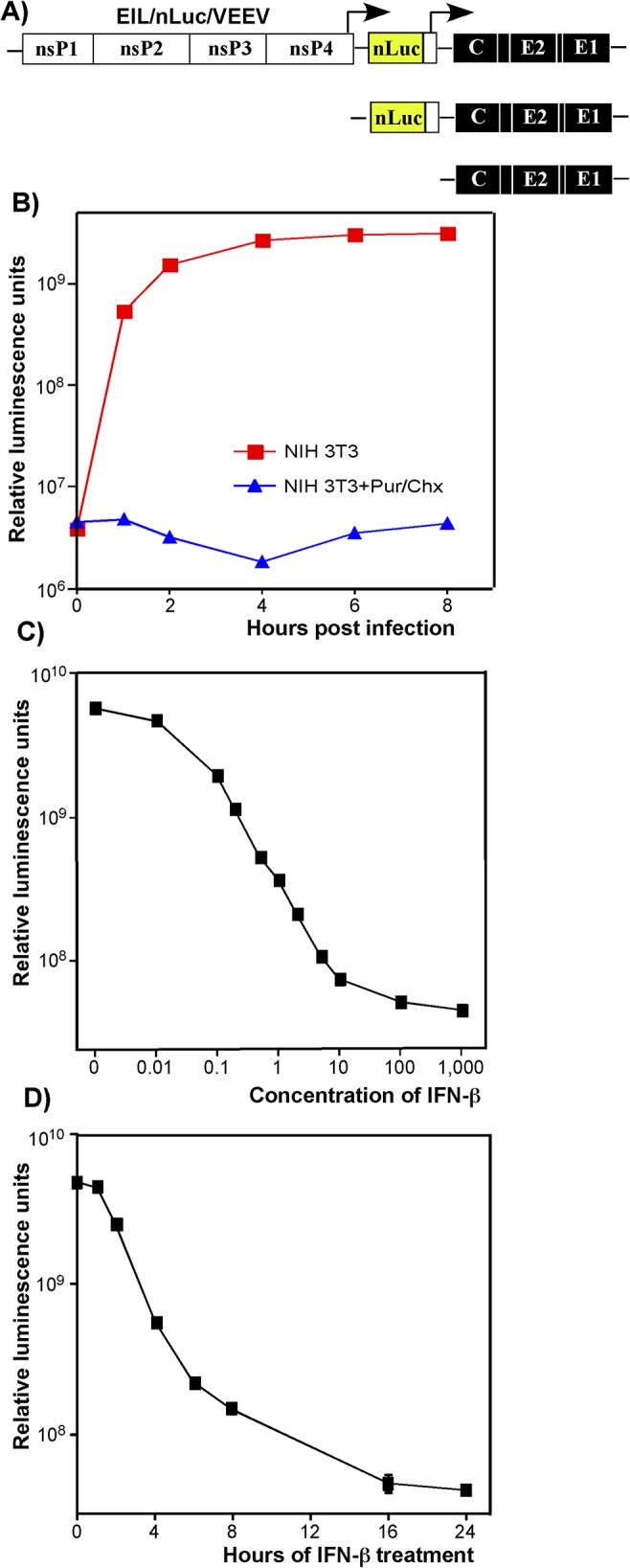
(A) The schematic representation of EIL/nLuc/VEEV genome and encoded SG RNAs. (B) NIH 3T3 cells were infected with purified EIL/nLuc/VEEV at an MOI of 10 PFU/cell. After 1 h incubation at 4°C, cells were washed with cold PBS, and then incubated at 37°C in complete media. At the indicated time points, cells were harvested, and nLuc activity was assessed. The control cells were infected and further incubated in the presence of 50 μg/ml puromycin (Pur) and 50 μg/ml cycloheximide (Chx). (C) NIH 3T3 cells were treated for 20 h with IFN-β at the indicated concentrations. Cells were then infected with EIL/nLuc/VEEV at an MOI of 10 PFU/cell for 1 h at 37°C, washed with PBS, and incubated in complete medium at 37°C. nLuc activity was assessed at 4 h post infection. (D) NIH 3T3 cells were treated for indicated times with IFN-β at a concentration of 100 IU/ml. Cells were then infected with EIL/nLuc/VEEV at an MOI of 10 PFU/cell, washed with PBS, and incubated in complete media at 37°C. nLuc activity was analyzed at 4 h post infection. All results are presented as average of triplicate +/- SD in relative luminescence units from a single representative experiment. Most of standard deviations are too small to be seen. The experiments were reproducibly repeated three independent times.