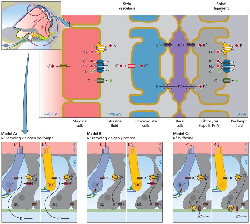
FIGURE 3. Overview over the stria marginalis and its K+ transport mechanisms and two alternative K+ pathways removing K+ from the hair cells.
The stria vascularis consists of three distinct cell types: marginal cells, intermediate, and basal cells. Intermediate cells are connected via gap junctions to basal cells, which in turn form connexons with underlying fibrocytes. Model A postulates that K+ released from hair cells cycles back to the stria vascularis through the open perilymph space, whereas model B entails K+ recycling through inner phalangeal cells, marked as supporting cells (SC) here, in the case of inner hair cells (IHC) or Deiters’ cells (DC) in the case of outer hair cells (OHC). These cells act as a K+ buffer in model C. Note that these three models are not mutually exclusive. Parts of this figure have been redrawn from Ref. 36a, with permission of the authors, editors, and publisher (Elsevier).

