
Fig. 2.
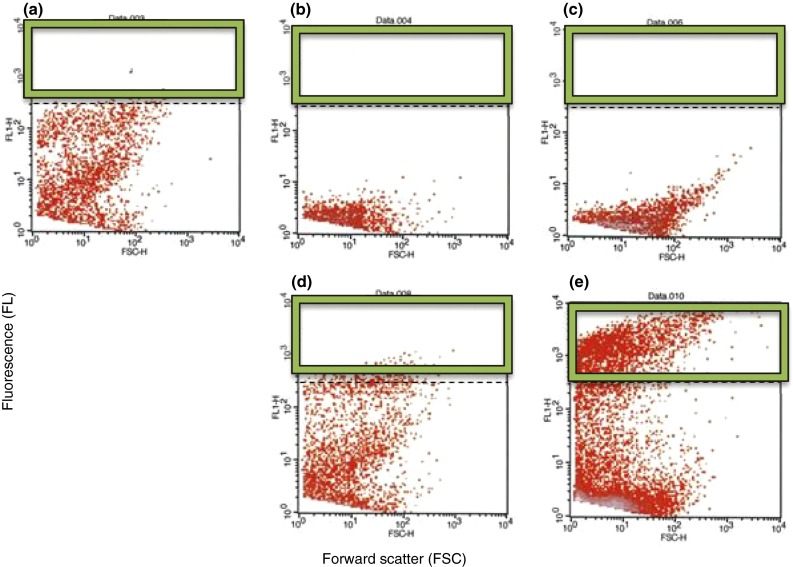
Co-aggregation of Lact. reuteri DSM17648 with H. pylori DSM21031 was analyzed by flow cytometry (e). H. pylori cells were CFDA stained. Samples were analyzed using flow cytometry, and cell co-aggregation was quantified by determining the events with a high FL (>5 × 10², area within green frame). Co-aggregation was not observed when strains were analyzed separately (a–c) nor when a non-aggregating Lactobacillus strain was used as a control (d)