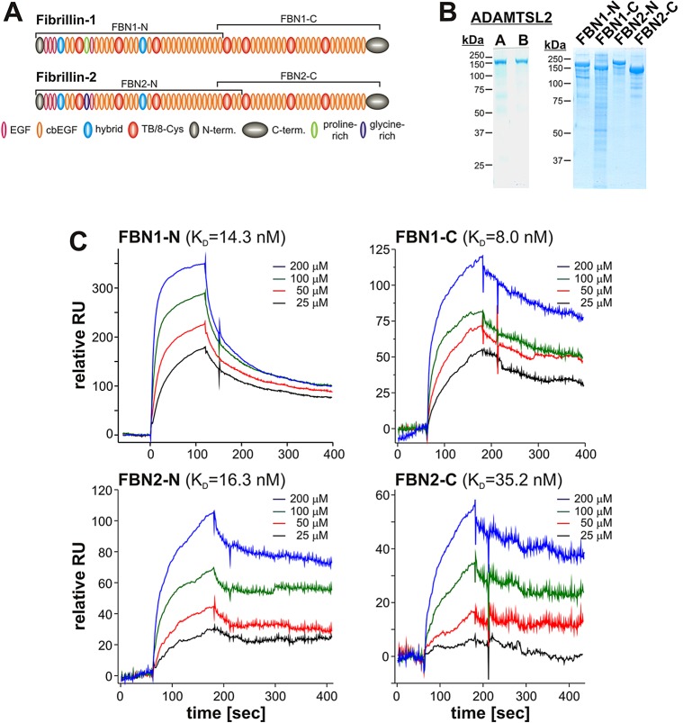
Fig. 7.
ADAMTSL2 binds to FBN1 and FBN2. (A) Domain structure of human FBN1 and FBN2. The peptides used in the binding assays with ADAMTSL2 are marked with brackets. (B) ADAMTSL2 (left, preparation A and B) and fibrillin peptides (right) were separated on 7.5% polyacrylamide gels under reducing conditions followed by Coomassie staining to demonstrate the purity of protein preparations. (C) Binding of immobilized ADAMTSL2 to soluble N- or C-terminal fragments of FBN1 or FBN2, respectively, was measured by surface plasmon resonance (Biacore) (n=2). The KD for FBN1-N was similar to that previously published for a slightly different N-terminal fragment (60 nM) (Le Goff et al., 2011).

