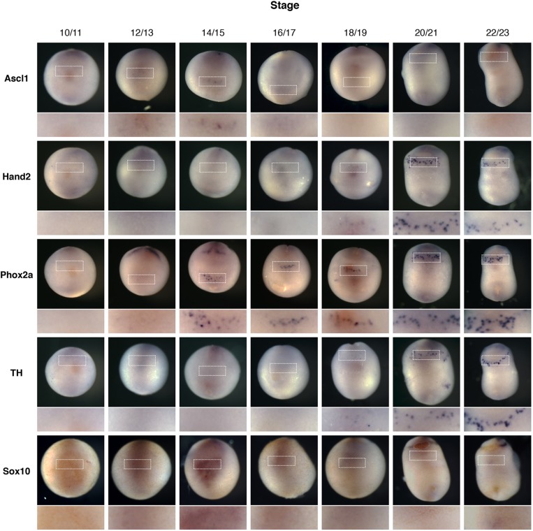
Fig. 2.
Ascl1 is expressed transiently during noradrenergic development in the anteroventral region. Xenopus embryos were fertilised and allowed to develop to the developmental stage indicated before fixing and staining by in situ hybridisation for noradrenergic (NA) neuron markers Ascl1, Hand2, Phox2a and TH, along with the neural crest marker Sox10. Embryos are all orientated with the ventral side imaged and head to the top. The anteroventral region where AVNA cell markers are expressed is expanded in the lower panel.

