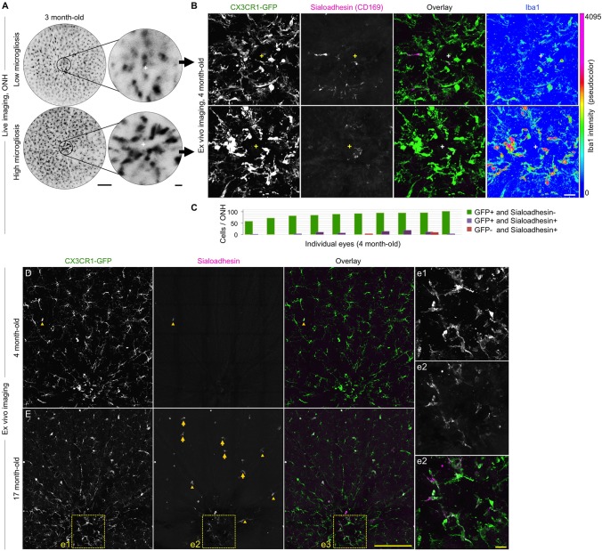
Fig. 6.
Early microgliosis is mainly driven by microglia resident in the ONH. (A) Live cSLO images of two 3-month-old retinas (left) and their ONHs (right, cross indicating its center), representative of low and high microgliosis. (B) Ex vivo confocal images of the same ONHs at 4 months of age, shown as maximal intensity projection of the inner 30 µm. Triple-immunostaining detects few cells that are positive for both GFP and sialoadhesin within the ONH, regardless of their different levels of microgliosis and activation, revealed by upregulated Iba1 expression in the ONH with increased microgliosis. (C) Number of cells expressing GFP and/or sialoadhesin per ONH area at 4 months of age. Single- and double-stained cells were quantified in confocal images spanning the central 200 µm of retinal whole mounts, throughout the inner 60 µm (0.8-µm z-slices). (D) Low magnification, single-slice view of the same 4-month-old retina representative of high microgliosis (B, bottom row). (E) Comparable view of a 17-month-old retina, showing sialoadhesin expression in perivascular (arrowheads), parenchymal (arrows) and ONH cells (frame). The ONH area is shown at higher magnification in corresponding insets. Scale bars: 250 µm (A,B,D and E), 25 µm (insets in A, and e).

