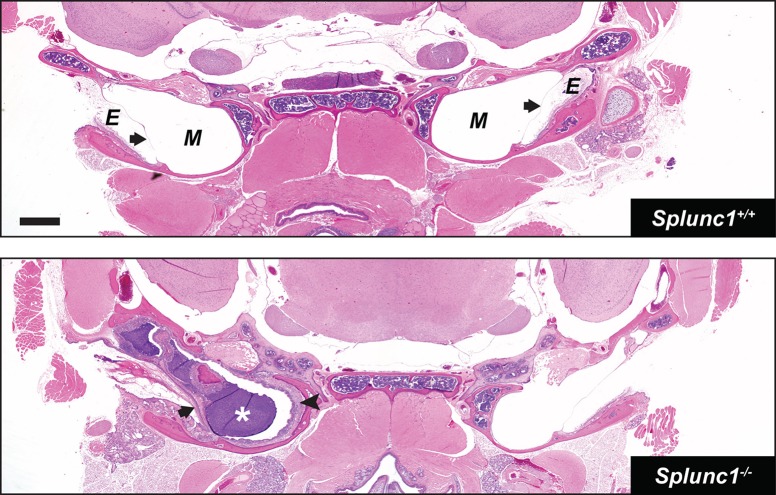
Fig. 2.
Unilateral otitis media in a Splunc1−/− mouse. H&E-stained coronal sections through the heads of 10- to 18-month-old Splunc1+/+ and Splunc1−/− mice were inspected for gross abnormalities. The top panel depicts a representative coronal section from a Splunc1+/+ mouse. The bottom panel is from a Splunc1−/− mouse exhibiting unilateral otitis media, characterized by purulent material in the middle ear lumen (white asterisk). In this mouse, thickening of the tympanic membrane and middle ear epithelium (black arrowhead) are indicative of chronic otitis media. E, external ear canal; M, middle ear; in both panels, black arrows indicate the tympanic membranes. Scale bar: 0.7 mm.

