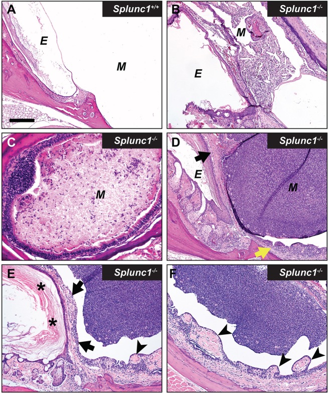
Fig. 3.
Otitis media in Splunc1−/− mice is incompletely penetrant. Panel A depicts the lumen of the middle ear from a representative Splunc1+/+ mouse, which is largely free of PMNs or epithelial hyperplasia. In contrast, middle ears from different Splunc1−/− mice (B-F) exhibited variable severity with respect to inflammation and epithelial remodeling at the time of necropsy. The abundance of accumulated PMNs and cell debris in the middle ear lumens of these mice ranged from moderate (B,C) to very significant (D-F). Occasionally, small spicule-shaped spaces known as cholesterol clefts (indicative of cholesterol crystals in the original tissue) were observed along with cellular debris in the middle ears of Splunc1−/− mice (B). Some Splunc1−/− mice also presented with varying amounts of epithelial proliferation and mucosal thickening in the middle ear (D, yellow arrow) and tympanic membrane (D, black arrow), suggesting a history of repeated otitis. In the mouse depicted in panels E and F, remodeling manifests as thickening of the tympanic membrane (black arrows) by hyperkeratosis (asterisks), inflammation and increased connective tissue. The region of middle ear adjacent to inflammation often had increased connective tissue, epithelial proliferation and/or polyploid changes (arrowheads). E, external ear canal; M, middle ear. Scale bar: 180 µm (A,B,D-F); 90 µm (C).

