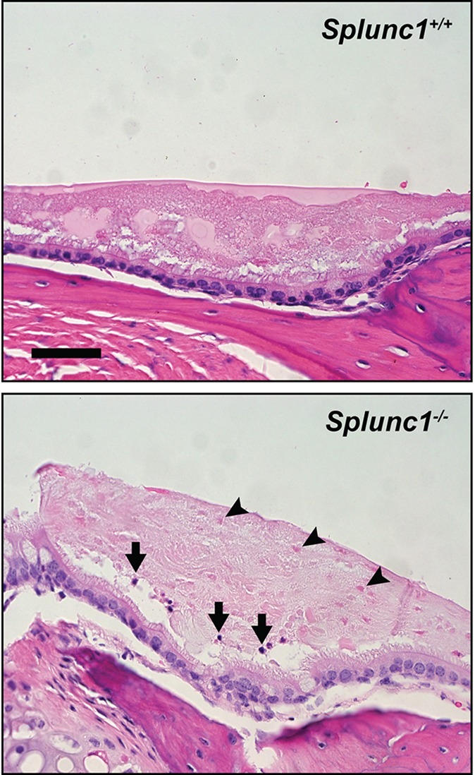
Fig. 4.

The middle ears of Splunc1−/− mice harbor increased inflammatory cells and cellular debris relative to wild-type littermate controls. The top panel depicts a representative image of a middle ear from a Splunc1+/+ mouse, in which the middle ear epithelium is covered by a layer of fluid containing globular fluid material. In the Splunc1−/− middle ear image (bottom panel) this fluid contains multiple punctate eosinophilic ‘ghost’ cells (black arrowheads) along with a small number of solitary PMNs (black arrows). Scale bar: 43 µm.
