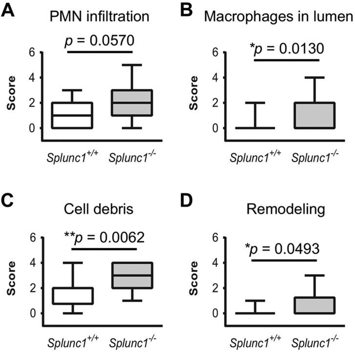
Fig. 7.
Summary of histopathological analysis of Splunc1−/− and wild-type mouse middle ears. Middle ear sections from Splunc1−/− mice and age-matched wild-type littermate controls were examined for histological evidence of otitis or other significant changes in the middle ear, using scoring rubrics described in the Materials and Methods. Sections were scored for several parameters including the infiltration of PMNs and macrophages in the middle ear lumen (panels A and B, respectively), accumulated cell debris (C), and the degree of hyperplasia and remodeling exhibited by the epithelium (D). Scores are summarized as box and whiskers plots. In these plots, the median score is denoted by a thick line, the box indicates the 25th and 75th percentiles, and the minimum and maximum values are depicted by the outer bars. Mann–Whitney tests were used to test for statistically significant differences (n=18 Splunc1+/+ and n=26 Splunc1−/− mice).

