Fig. 1.
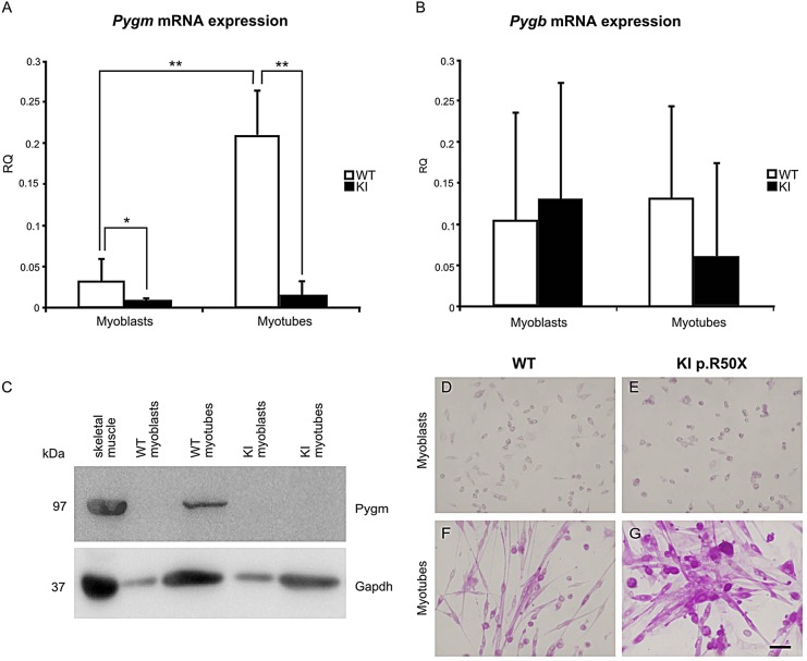
Differential glycogen phosphorylase expression in mouse primary skeletal-muscle cultures. Only WT myotubes expressed Pygm mRNA (A). Myoblasts and myotubes from WT and KI mice expressed Pygb mRNA with a high variation and no statistically significant differences between WT and KI (B). Presence of both Pygm transcript and protein (GP-MM) was observed only in WT myotubes (C). PAS staining of WT myoblasts (D), WT myotubes (F), KI myoblasts (E) and KI myotubes (G): only KI myotubes accumulated high glycogen levels (G). KI, knock-in; WT, wild type; RQ, relative quantification. *P<0.05 for the comparison KI versus WT, **P<0.001 for the comparison of KI versus WT. Scale bar: 50 µm.

