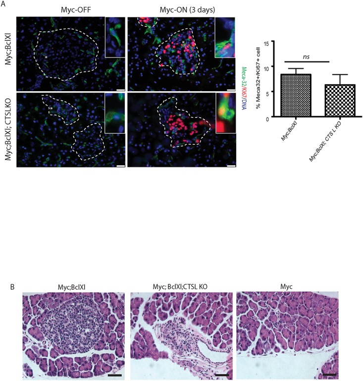
Fig 3. Loss of cathepsin L does not inhibit onset of Myc-induced tumorigenesis.

(A) Immunohistochemical analysis of endothelial cell proliferation in vivo. Pancreata was isolated from the MycER TAM ;Bcl-xL animals from CTS L WT and CTS L-deficient backgrounds untreated or treated with TAM for the duration of three days. Proliferating endothelial cells were identified by co-labeling with the endothelial marker Meca-32 (green) and Ki67 (red). The islet area is outlined by dotted lines. The asterisks indicate the magnified areas of the endothelial compartment of islets presented in the insets. The percentage of Meca-32-positive cells that also stained positive for the proliferation marker Ki67 was then determined as described in Materials and Methods section. At least three animals were assayed of each genotype all analyses done in duplicate; ten randomized fields per analysis were considered. The graph shows the mean and standard error of the mean. ns—no statistical significant difference was detected by Student’s T-test analysis. Scale bars, 25μm. (B) Representative H&E staining of pancreatic sections from MycER TAM, MycER TAM ;Bcl-xL and MycER TAM ;Bcl-xL;CTSLKO collected from animals subjected to Myc activation for 3 days. Induction of Myc in the pancreatic islets lacking Bcl-xL expression (Myc) induces profound apoptosis and ablation of the islet beta-cell compartment as described in [28] (right panel). Loss of cathepsin L in MycER TAM ;Bcl-xL;CTSLKO (middle panel) has no significant impact on morphology of pancreatic islets following onset of Myc-induced tumorigenesis when compared to MycER TAM ;Bcl-xL islets (left panel). At least three animals of each genotype were assayed, eight randomized fields per analysis were considered. Scale bars, 25μm.
