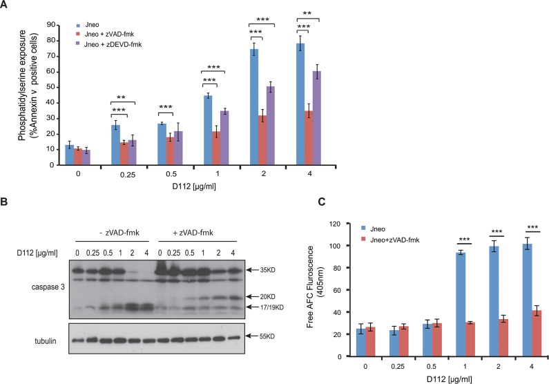
Fig 2. D112 induces caspase activation in Jurkat cells.
(A) Phosphatidylserine exposure. Jneo cells were treated with indicated concentrations of D112 for 24 h in the presence or absence of the caspase 3 inhibitor z-DEVD-fmk (20 μM), stained as indicated and analyzed by flow cytometry. Mean ± SD of three independent experiments performed in triplicate are shown, *P<0.05, **P<0.01, ***P<0.001. (B) Caspase cleavage. Cells were incubated for 24 h with increasing concentrations of D112 as indicated. Whole cell lysates were subjected to western blot analysis with the indicated antibodies. The experiment was performed independently three times and a representative blot is shown. (C) Caspase 3 enzymatic activation. Jneo cells were treated with D112 in the presence or absence of z-VAD-fmk (20 uM) for 24 h prior to lysis. Cell lysates were incubated with the caspase 3 specific fluorometric substrate, Ac-DEVD-AFC, for 1 h and caspase 3 activity was measured at 405 nm. Mean ± SD of three independent experiments performed in triplicate are shown, *P<0.05, **P<0.01, ***P<0.001.

