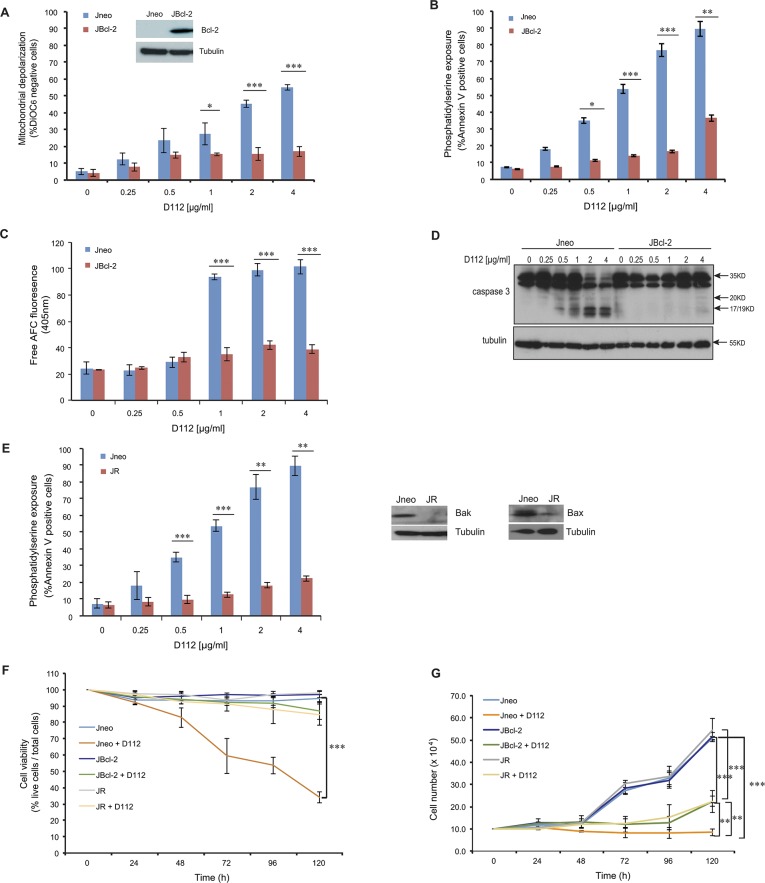
Fig 3. D112-induced apoptosis is blocked by Bcl-2.
Jneo and JBcl-2 cells were treated with the indicated amounts of D112 for 24 h. Apoptotic cell death was measured by (A) mitochondrial dysfunction, (B) phosphatidylserine exposure, (C) caspase 3 enzymatic activation, and (D) caspase cleavage. All of these apoptotic hallmarks were quantitated as described previously. (E) Bax/Bak expression is required for D112-induced cell death. Jneo and JR cells were treated with indicated concentrations of D112 for 24 h and phosphatidylserine positivity was recorded as done previously. (F) Inhibition of the apoptosis pathway protects cell from D112-induced cell death. Cells were treated with 62.5 ng/ml D112 for 24 h. Cells then were incubated in fresh medium and counted daily. Cell survival was detected by trypan blue staining. (G) Cell proliferation is inhibited by D112. Cells were treated with 62.5 ng/ml D112 for 24 h. Cells then were incubated in fresh medium and total live cell number was counted. The expression of Bcl-2, Bax and Bak were verified in western blot insets in A and E. In all experiments, the mean ± SD of three independent experiments performed in triplicate are shown, *P<0.05, **P<0.01, ***P<0.001.

