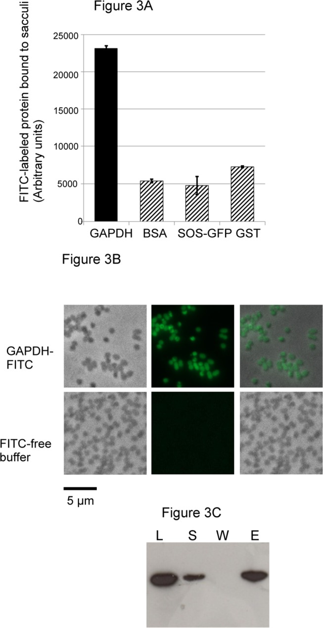
Fig 3. Pneumococcal GAPDH binds to the cell wall.

(A) Solid-phase binding assay of FITC-labeled proteins to pneumococcal cell wall sacculi containing the peptidoglycan and the teichoic acids. (B) Microscopic images of FITC-labeled GAPDH and FITC-free buffer used a negative control bound to pneumococci cell wall sacculi. Phase contrast, fluorescence and merge pictures are shown. Scale bars, 5 μm. (C) Pull down of GAPDH with pneumococcal cell wall. The load (L) protein was mixed with the insoluble cell wall preparation. The supernatant (S) fraction containing unbound protein was recovered. After extensive wash (W), protein bound to the cell wall pellet was eluted with Laemmli buffer at 100°C for 10 min (E). The protein samples were analyzed by Western blot using an anti-His tag antibody.
