Fig. 2.

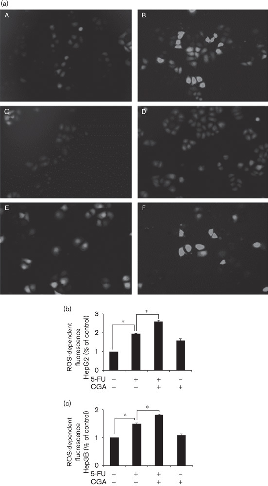
CGA increased 5-FU-induced ROS production in hepatocellular carcinoma cell. HepG2 cells were treated with control (A), 3% H2O2 as the positive control (B), 125 μmol/l CGA (C), 250 μmol/l CGA (D), 20 μmol/l 5-FU (E), and a combination of 20 μmol/l 5-FU and 250 μmol/l CGA (F) for 24 h. (a) Intracellular ROS production was detected using the ROS detection probe CM-H2DCFDA. The fluorescence intensity of ROS was directly measured using fluorescence microscopy (magnification, ×200). (b) Intracellular ROS production in HepG2 cells treated with 20 μmol/l 5-FU, 250 μmol/l CGA, and the combination of 20 μmol/l 5-FU and 250 μmol/l CGA was quantified using flow cytometric analysis. (c) Intracellular ROS production in Hep3B cells treated with 20 μmol/l 5-FU, 250 μmol/l CGA, and the combination of 20 μmol/l 5-FU and 250 μmol/l CGA was also quantified using flow cytometric analysis. Experiments were conducted in triplicate. Data are shown as the mean±SEM (n=3). *P<0.05 compared with the control. CCK-8, cell counting kit-8; CGA, chlorogenic acid; CM-H2DCFDA, 5-(and-6-)chloromethyl-2′,7′-dichlorodihydrofluorescein diacetate; 5-FU, 5-fluorouracil; ROS, reactive oxygen species.
