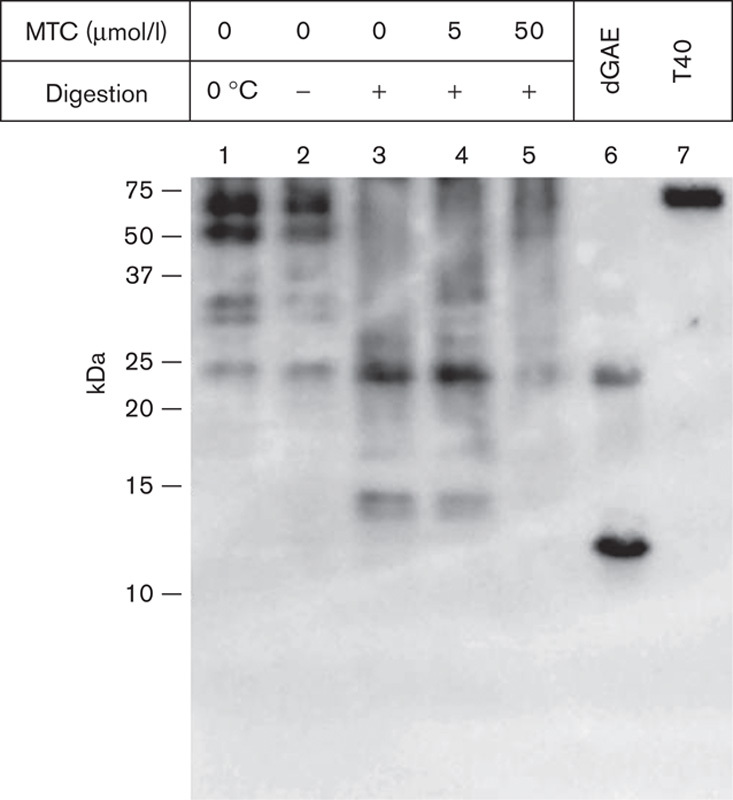
Fig. 10.

MTC disrupts tau aggregates extracted from mouse brains. Insoluble tau fractions were prepared from the brains of 8-month-old L66 mice that had not been treated with drug. These fractions were prepared using the method to produce non-Pronase PHFs from AD tissue. Fractions were incubated with micrococcal nuclease (2 U/ml in the presence of 3 mmol/l MgCl2) for 1 h at 37°C and centrifuged for 30 min at 42 000g; the pellet was analysed by SDS-PAGE and by immunoblotting with mAb 7/51. This led to the generation of a core-tau fragment having the gel mobility of a 12/14 kDa fragment (lane 3) and a more prominent dimer. These were decreased in intensity or absent when MTC was included at 5 and 50 μmol/l (lanes 4 and 5). Control samples were incubated for 1 h either on ice (lane 1) or at 37°C (lane 2) in the absence of nuclease. Tau proteins were included for reference: recombinant, truncated tau297–391 (dGAE; Novak et al., 1993) expressed in Escherichia coli and full-length htau40 (T40) expressed in eukaryotic cells. The positions of molecular weight markers, in kDa, are shown on the left. AD, Alzheimer’s disease; L66, Line 66; mAb, monoclonal antibody; MTC, methylthioninium chloride.
