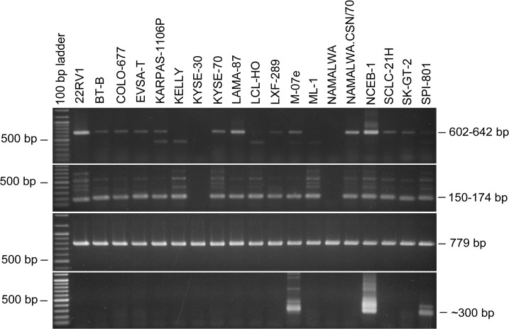
Fig 1. PCR-Assays for the sensitive and reliable detection of MLV sequences in cell line DNA.
The upper two panels show the ethidium bromide stained gels of the MLV-specific PCR assays performed with the outer (first panel) and inner primers (second panel). The sizes of the MLV-specific bands are 604–642 bp and 150–174 bp, respectively, depending on the MLV genotype. Some cell lines produce an unspecific band with the outer primers (also seen in some MLV-negative cell lines). The inner primers usually also produce several unspecific bands seen only in MLV-positive cell lines. The cell lines KYSE-30 and NAMALWA are MLV-negative in the assay, whereas the cell lines from the same series or subclones are MLV-positive, respectively. The third panel demonstrates the integrity of the DNAs used for the analyses by amplification of the human ABL gene. The lower panel shows contamination of the genomic DNA with mouse DNA by amplification of mouse specific IAP coding sequences. The PCR produces a double band of approximately 300 bp. The cell line NCEB-1 harbors several mouse-derived chromosomes.

