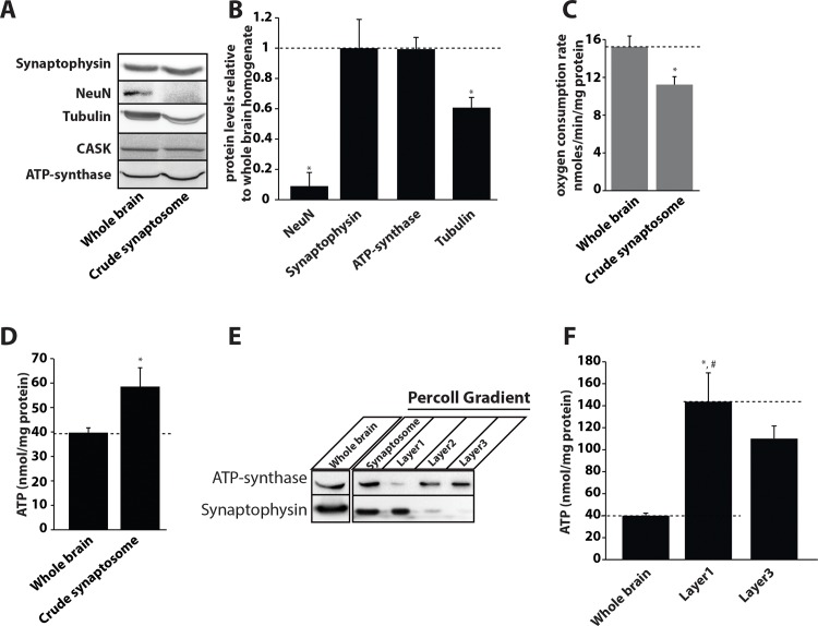
Fig 5. Neuronal presynapses are highly enriched in ATP.
A) Immunoblots from whole brain and crude synaptosomal fractions detected with the specified antibodies. B) Relative protein quantification from three different synaptosomal preparations compared to the whole brain. (Absolute value for whole brain is depicted as 1). Data are plotted as mean ± SEM. C) Mitochondrial oxygen consumption rate in crude synaptosomes and whole brain homogenates. Total oxygen consumption was normalized to the protein levels in both fractions. D) Total ATP levels in crude synaptosomes and whole brain normalized to the protein concentrations in each fraction. E) Percoll density gradient separation of mitochondrial and synaptosomal fractions. Synaptophysin was used as a marker for synaptosomes and ATP synthase β-subunit was used as a marker for mitochondria. F) Total ATP levels detected in the whole brain homogenate, synaptosomal and mitochondrial membrane-rich fractions obtained from the Percoll density gradient method. Data were normalized to total protein levels in each fraction and plotted as mean ± SEM.

