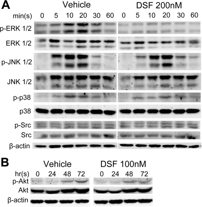
Fig 4. DSF attenuates RANKL-induced MAPK signaling in BMMs.

Total cell lysates were extracted from BMMs treated with rRANKL for 0, 5, 10, 20, 30 and 60 mins (A) or for 0, 24, 48 and 72 hrs (B) in the presence or absence of DSF (200nM or 100nM). Proteins were separated on 12.5% SDS-PAGE gel, transferred onto nitrocellulose membranes, and immunoblotted sequentially with antibodies to different components of the MAPK and SAPK signaling pathways (ERK, JNK, p38, Src and Akt). β-actin was used as internal loading control. Results shown represent one of three independent experiments.
