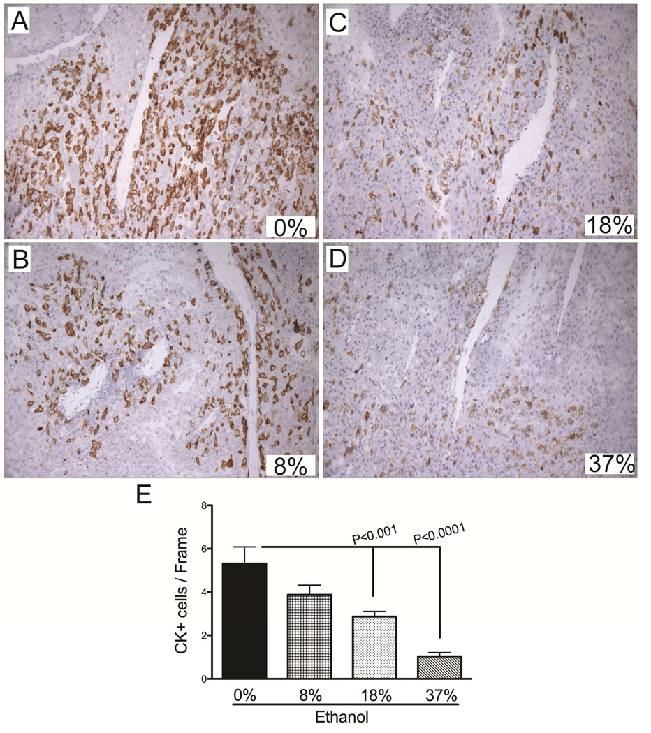
Figure 3.
Gestational ethanol exposure reduces invasive trophoblasts at the implantation site. A–D) Cytokeratin (CK) immunostained sections demonstrate the distribution of invasive trophoblasts at the mesometrium of dams exposed to 0%, 8%, 18%, or 37% of ethanol by caloric content. Invasive trophoblasts were quantified by stereology. Graph (E) depicts the mean ± S.E.M. corresponding to the numerical density of CK-positive cells in each group. Inter-group comparisons were made using one-way ANOVA with the Dunnett’s multiple comparison test.

