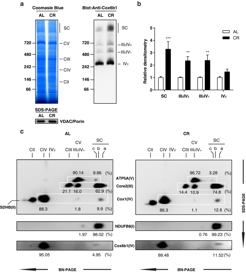
Fig. 2.

CR promotes the assembly of mitochondrial supercomplexes. a Blue native polyacrylamide gel electrophoresis (BN-PAGE) of liver tissue samples from 12-month-old mice fed AL or 30 % CR. Coomassie blue staining of 1D BN-PAGE gel (left panel) and subsequent immunoblotting with anti-Cox6b1 antibody (right panel). SDS-PAGE and immunoblotting with anti-VDAC/Porin antibody was also performed to confirm equivalent protein loading of mitochondria lysates (lower panel). b Densitometric analysis of immunoblots. The results are presented as mean ± SD (n = 6, **p < 0.01, ***p < 0.001, vs. AL group). c 2D BN/SDS-PAGE of AL and CR liver samples. Immunoblotting was performed using the anti-ATP5A antibody for Complex V, the anti-Core2 antibody for Complex III, the anti-SDHB antibody for Complex II, and the anti-Cox1 and the anti-Cox6b1 antibodies for Complex IV. The position SC-a corresponds to the mitochondrial supercomplexes containing I/III2/IVn (n = 1, 2, 3, or 4); the position SC-b corresponds to I/III2; The position SC-c corresponds to Complex V dimers. The positions of Complex V monomer (CV), Complex III monomer (CIII), Complex III dimers + Complex IV monomers or dimers (III2IVn, n = 1 or 2), Complex IV monomer or dimers (CIV or IV2), and Complex II monomers (CII) are also indicated. CV and III2IVn spots are overlapping. The values represent the proportion of signal intensity in each spot in a row detected by a specific antibody
