Figure 1.
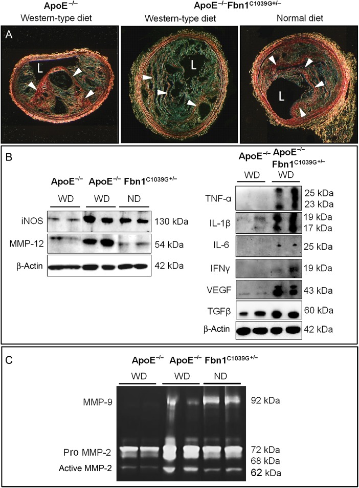
Atherosclerotic plaques in brachiocephalic arteries of ApoE−/−Fbn1C1039G+/− mice on Western-type diet show features of a highly unstable phenotype when compared with age-matched ApoE−/− mice on Western-type diet and ApoE−/−Fbn1C1039G+/− mice on normal diet (20 weeks of diet on average). (A) type I collagen (red/arrowheads, sirius red stain under polarized light) was decreased fivefold (for quantification, see Supplementary material online, Table S1). Scale bar = 100 µm. L, lumen. (C) Inducible nitric oxide synthase expression was augmented in plaques of ApoE−/−Fbn1C1039G+/− mice on Western-type diet and normal diet when compared with ApoE−/− mice on Western-type diet, indicating increased macrophage activation. Accordingly, inflammatory cytokines TNF-α, IL-1β, and IL-6 were highly expressed in aortas of ApoE−/−Fbn1C1039G+/− mice on Western-type diet compared to ApoE−/− mice. In addition, an increase in T-cell activation marker interferon γ, vascular endothelial growth factor, transforming growth factor-β, and elastase MMP-12 was observed. (D) Gel zymography showed increased activity of MMP-9 and MMP-2 when compared with aortas of age-matched ApoE−/− mice on Western-type diet. For quantification, see Supplementary material online, Figure S2.

