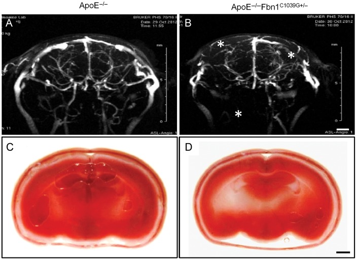
Figure 4.
Brains of ApoE−/−Fbn1C1039G+/− mice on Western-type diet displayed disturbed cerebral flow and hypoxia, indicative of stroke. (A and B) Representative cerebral MR angiograms of ApoE−/− (A) and ApoE−/−Fbn1C1039G+/− mice (B) on Western-type diet for 25 weeks. ApoE−/−Fbn1C1039G+/− mice showed perfusion deficits in both right and left brain hemispheres (asterisks), in comparison to age-matched ApoE−/− mice (A) (n = 11 and n = 7, respectively, P < 0.001, Fisher's exact test). (C and D) Representative 2,3,5-triphenyltetrazolium chloride stain of brains of an ApoE−/− and ApoE−/−Fbn1C1039G+/− mouse on Western-type diet for 25 weeks. Vital brain tissue is stained red, while hypoxic areas are pale/white. Sixty-four percent of ApoE−/−Fbn1C1039G+/− mice showed hypoxic areas in the brain, indicative of stroke (D; white/pale regions, arrowheads, n = 11), whereas brains of most ApoE−/− mice on Western-type diet (C) were normal (87%, n = 8) (P < 0.001, Fisher's exact test). Scale bar = 200 µm.

