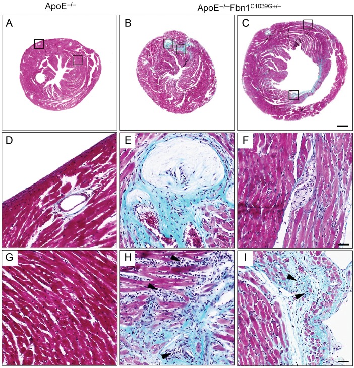
Figure 5.
Sections of hearts of an ApoE−/− mouse at 35 weeks on Western-type diet and two ApoE−/−Fbn1C1039G+/− mice that died suddenly (Masson's trichrome stain). Hearts of ApoE−/−Fbn1C1039G+/− mice showed large infarcted areas, mainly in the left ventricle (green-blue, B, C and H, I) and coronary artery plaque (B, C and E, F), whereas hearts of ApoE−/− mice appeared normal (A, D, G). Scale bar = 500 µm. (E, F) Detail of coronary artery plaque and infarcted area (H and I) in B and C, respectively, in the left ventricle. Infarcted areas showed infiltration of inflammatory cells (arrowheads). The corresponding areas in ApoE−/− mice on Western-type diet appeared normal and coronary arteries were plaque free (D, G). Scale bar = 50 µm.

