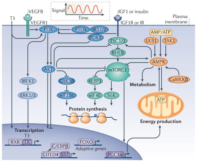
Figure 1. Physiological hypertrophy signalling pathways.
Physiological hypertrophy is an adaptive form of cardiac hypertrophy. The figure depicts central signalling pathways of physiological hypertrophy discussed in the text. Physiological hypertrophy is initiated by intermittent signals of triiodothyronine (T3), vascular endothelial growth factor B (VEGFB), insulin and insulin-like growth factor 1 (IGF1), as illustrated by the oscillating curve. The growth hormones activate membrane-localized tyrosine kinase receptors (VEGF receptor 1 (VEGFR1), IGF1 receptor (IGF1R) or insulin receptor (IR)) and nuclear receptors (thyroid hormone receptor (TR)), which trigger intracellular signalling pathways specific to physiological hypertrophy. These signalling pathways regulate the transcription of adaptive genes, protein synthesis, metabolism and energy production. The growth signals centre on common signalling branches controlled by ERK1/2, PI3K, AKT and mTOR complex 1 (mTORC1), whereas AMP-activated protein kinase (AMPK) governs metabolic adaptive reprogramming. 4EBP1, eukaryotic translation initiation factor 4E-binding protein 1; C/EBPβ, CCAAT/enhancer binding protein-β; CaMKKβ, calcium/calmodulin-dependent protein kinase kinase-β; CITED4, CBP/p300 interacting transactivator 4; eIF2Bε, eukaryotic translation initiation factor 2Bε; FOXO, forkhead box O; GSK3β, glycogen synthase kinase 3β; IRS1/2, insulin receptor substrate 1 or 2; LKB1, liver kinase B1; PDK1, phosphoinositide-dependent protein kinase 1; PGC1α, peroxisome proliferator-activated receptor-γ co-activator 1α; RHEB, RAS homologue enriched in brain; RXR, retinoic acid receptor; S6K, S6 kinase; SRF, serum response factor; TAK1, transforming growth factor β-activated kinase 1; TSC1/2, tuberous sclerosis complex 1 or 2.

