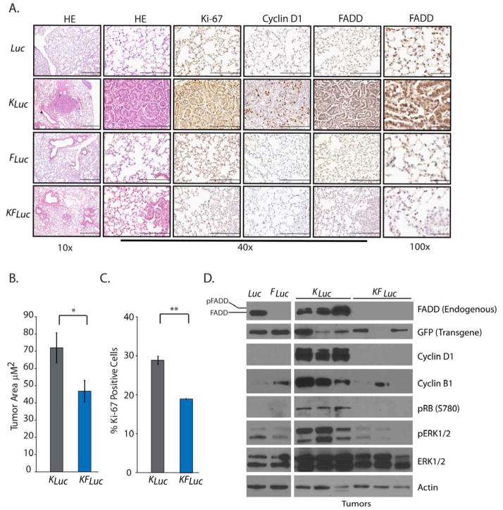
Fig. 2. Fadd null lung tumors are less proliferative.
(A) Representative histology images of lungs removed from mice 18 weeks after AdCre administration. Tissues were stained with hematoxylin and eosin (HE) or antibodies against cyclin D1, Ki-67, and FADD. Scale bars: 10x, 500 μm; 40x, 200 μm; 100x, 50 μm. (B) Average tumor area quantified from H&E-stained lung tissue sections from KLuc (n=6) and KFLuc (n=10) mice. *P=0.04, unpaired Student’s t-test. (C) Average percentage of positive Ki-67 stained cells in lung tissue sections from KLuc and KFLuc mice, 4 fields per 10 mice each. **P=2×10⁻⁵, unpaired Student’s t-test. (D) Representative Western blot for endogenous FADD, GFP (Fadd transgene), phosphorylated and total ERK1/2, phosphorylated RB, cyclin D1, cyclin B1 and β-Actin in lung tissue from the indicated mice. Blots are representative of 3 independent experiments.

