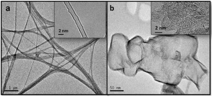
Figure 1.

Representative transmission electron microscopy micrographs of the graphene and single-walled carbon nanotube nanomaterials used in this study. (a) Transmission electron microscopy micrographs of the (a) crystalline carboxylated single-walled carbon nanotube, and (b) carboxylated nano-graphene structures synthesized on the Fe:Mo:MgO bimetallic system. Each inset shows the same material at a higher resolution.
