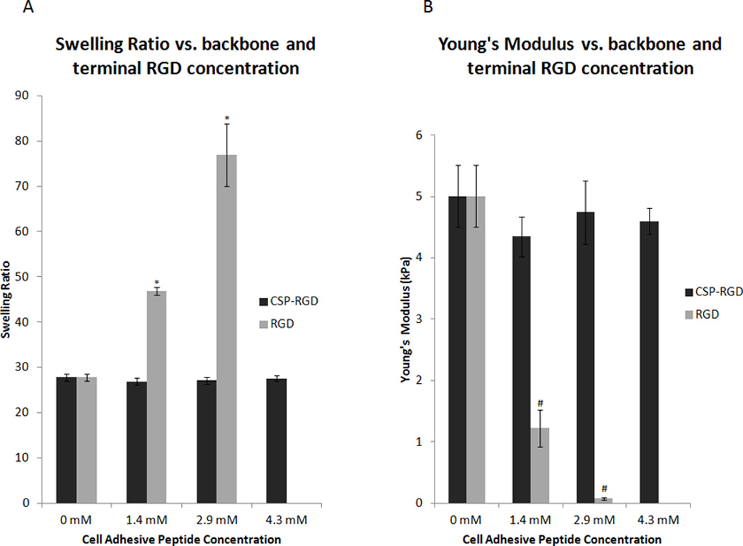
Figure 3.

(A) Average swelling ratios for four-arm PEG hydrogels (10% w/v) with CSP-RGD (black bars) and RGD (grey bars) peptide concentrations of 0 mM, 1.4 mM, 2.9 mM, and 4.3 mM. CSP-RGD represents hydrogels where the cell adhesive peptide was attached to the backbone of the polymer network, whereby RGD refers to hydrogels that had the cell adhesive peptide attached to terminal ends of PEG. Hydrogels containing the combination CSP-RGD sequence displayed no significant change in swelling ratio with respect to concentration of the combination peptide. Hydrogels containing the RGD sequence possessed significant increases in swelling ratio with respect to the RGD peptide concentration. *: p<0.05 compared to CSP-RGD hydrogels with the same cell adhesive peptide concentration. (B) Young’s Modulus for four-arm PEG hydrogels (10% w/v) with CSP-RGD concentrations of 0 mM, 1.4 mM, 2.9 mM, and 4.3 mM were 5.0 ± 0.5, 4.3 ± 0.3, 4.7 ± 0.5, and 4.6 ± 0.2 kPa respectively. Young’s Modulus for four-arm PEG hydrogels (10% w/v) with these same concentrations of RGD peptide were 5.0 ± 0.5, 1.2 ± 0.3, 0.1 ± 0.02 and no hydrogel formed respectively. The force applied to the hydrogels ranged from 8 mN to 10 mN. There was no significant difference in the Young’s Modulus of any of the hydrogel samples when the RGD sequence was linked to the backbone of the hydrogel network, however, terminally linked RGD resulted in a significant change in Young’s Modulus. #: p < 0.05 compared to CSP-RGD hydrogels with the same cell adhesive peptide concentration.
